 Back
BackStudy Guide: Kidney Anatomy, Blood Flow, and Nephron Structure
Study Guide - Smart Notes
 Tailored notes based on your materials, expanded with key definitions, examples, and context.
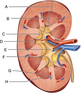
Q1. Assign the letters (A–H) to their corresponding regions and structures shown in the figure of the internal anatomy of the kidney.
Background
Topic: Kidney Anatomy
This question tests your knowledge of the gross anatomical regions and structures of the kidney, which are essential for understanding renal function and urine formation.
Key Terms:
Renal cortex
Renal medulla
Renal pyramid
Renal column
Renal pelvis
Major and minor calyces
Papilla of pyramid
Step-by-Step Guidance
Examine the labeled diagram and identify the outermost region of the kidney, which is typically the renal cortex.
Locate the region just beneath the cortex, which is the renal medulla, containing the renal pyramids.
Identify the triangular structures within the medulla—these are the renal pyramids.
Find the area between the pyramids, which is the renal column.
Trace the pathway of urine from the papilla of the pyramid into the minor calyx, then into the major calyx, and finally into the renal pelvis.
Try solving on your own before revealing the answer!
Final Answer:
A: Renal cortex B: Renal medulla C: Major calyx D: Papilla of pyramid E: Renal pelvis F: Minor calyx G: Renal pyramid H: Renal column
Each label corresponds to a distinct anatomical region or structure within the kidney, which plays a role in urine formation and transport.
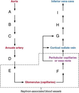
Q2. Assign the letters (A–I) to the missing blood vessels in the path of blood flow through the kidney.
Background
Topic: Renal Blood Flow
This question assesses your understanding of the sequence of blood vessels that supply and drain the kidney, which is crucial for filtration and renal function.
Key Terms and Pathway:
Renal artery
Segmental artery
Interlobar artery
Cortical radiate artery
Afferent arteriole
Glomerulus
Efferent arteriole
Arcuate vein
Interlobar vein
Renal vein
Step-by-Step Guidance
Start with the renal artery, which branches from the aorta and enters the kidney.
Follow the sequence: renal artery → segmental artery → interlobar artery → arcuate artery → cortical radiate artery.
Identify the afferent arteriole, which leads to the glomerulus for filtration.
After filtration, blood exits via the efferent arteriole and enters peritubular capillaries or vasa recta.
Trace the venous return: cortical radiate vein → arcuate vein → interlobar vein → renal vein → inferior vena cava.
Try solving on your own before revealing the answer!
Final Answer:
A: Renal artery B: Segmental artery C: Interlobar artery D: Cortical radiate artery E: Afferent arteriole F: Efferent arteriole G: Arcuate vein H: Interlobar vein I: Renal vein
This sequence ensures efficient filtration and reabsorption in the kidney.
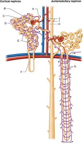
Q3. Assign the letters (A–J) to the corresponding parts of a nephron and associated structures.
Background
Topic: Nephron Anatomy
This question tests your ability to identify the structural components of a nephron, the functional unit of the kidney, and their associated blood vessels.
Key Terms:
Glomerulus
Glomerular capsule (Bowman's capsule)
Proximal convoluted tubule (PCT)
Nephron loop (Loop of Henle)
Distal convoluted tubule (DCT)
Collecting duct
Afferent and efferent arterioles
Peritubular capillaries
Vasa recta
Step-by-Step Guidance
Identify the glomerulus and glomerular capsule, which together form the renal corpuscle.
Trace the path of filtrate: from the glomerular capsule to the proximal convoluted tubule, then through the nephron loop, and into the distal convoluted tubule.
Recognize the collecting duct, which receives filtrate from multiple nephrons.
Locate the afferent and efferent arterioles, which supply and drain the glomerulus.
Identify the peritubular capillaries and vasa recta, which are involved in reabsorption and maintaining the medullary osmotic gradient.
Try solving on your own before revealing the answer!
Final Answer:
A: Efferent arteriole B: Glomerulus C: Glomerular capsule D: Proximal convoluted tubule E: Peritubular capillaries F: Nephron loop G: Vasa recta H: Distal convoluted tubule I: Collecting duct J: Afferent arteriole
Each structure plays a specific role in filtration, reabsorption, and secretion within the nephron.
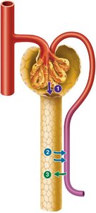
Q4. Identify the three major renal processes (labeled 1–3) indicated on the nephron diagram.
Background
Topic: Renal Physiology
This question focuses on the three key processes of urine formation: glomerular filtration, tubular reabsorption, and tubular secretion.
Key Terms:
Glomerular filtration
Tubular reabsorption
Tubular secretion
Step-by-Step Guidance
Examine the diagram and locate the glomerulus, where filtration occurs (process 1).
Identify the renal tubule, where reabsorption of water and solutes back into the blood takes place (process 2).
Find the areas where substances are secreted from the blood into the filtrate (process 3).
Try solving on your own before revealing the answer!
Final Answer:
1: Glomerular filtration 2: Tubular reabsorption 3: Tubular secretion
These processes are essential for the formation and composition of urine.
