 Back
Study Guide: Lower Appendage Bones – Structure and Function
Study Guide - Smart Notes
 Tailored notes based on your materials, expanded with key definitions, examples, and context.
Lower Appendage Bones
Overview of the Lower Appendage Skeleton
The lower appendage skeleton consists of the bones of the pelvis, thigh, leg, ankle, and foot. These bones provide support, facilitate movement, and protect internal organs. Understanding their structure and function is essential for anatomy and physiology students.
The Pelvic Girdle
Structure of the Hip Bone (Os Coxa)
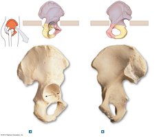
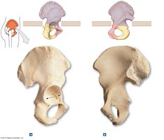
The hip bone, or os coxa, is composed of three fused bones: the ilium, pubis, and ischium. It forms the pelvic girdle, which connects the lower limb to the axial skeleton.
Ilium: The broad, superior portion of the hip bone.
Pubis: The anterior, inferior part of the hip bone.
Ischium: The posterior, inferior part of the hip bone.
Acetabulum: The deep socket that articulates with the head of the femur.
Obturator foramen: Large opening for nerves and blood vessels.
Iliac crest: The superior border of the ilium.
Pelvic Landmarks and Divisions
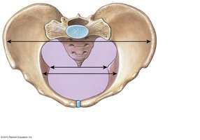
The pelvis is divided into the false pelvis and true pelvis, with important landmarks for anatomical orientation and childbirth.
Pelvic brim: The edge separating the false and true pelvis.
Pelvic inlet: The upper opening of the true pelvis.
Pelvic outlet: The lower opening of the true pelvis.
Arcuate line: Part of the pelvic brim.
Pubic symphysis: Cartilaginous joint uniting the left and right pubic bones.
Sex Differences in the Human Pelvis
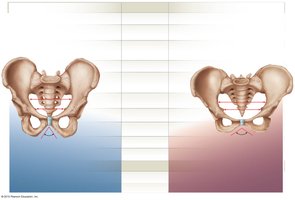
The human pelvis exhibits distinct differences between males and females, primarily related to childbirth. These differences include the shape, size, and orientation of pelvic bones and openings.
Feature | Female Pelvis | Male Pelvis |
|---|---|---|
General Appearance | Broader, smoother, less robust | Narrower, rougher, more robust |
Pelvic Inlet | Open, circular shaped | Narrower, heart shaped |
Pelvic Outlet | Enlarged | Narrow |
Pubic Angle | 100º or more | Under 90º |
Bone Weight | Lighter | Heavier |
Bone Markings | Less prominent | More prominent |
Femur: The Thigh Bone
Structure and Bone Markings of the Femur
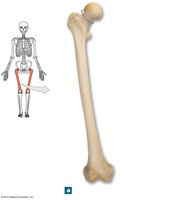
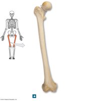
The femur is the longest and strongest bone in the human body, forming the thigh. It articulates proximally with the pelvis at the acetabulum and distally with the tibia and patella.
Head: Articulates with the acetabulum.
Fovea capitis: Small pit on the head for ligament attachment.
Neck: Connects the head to the shaft.
Greater and Lesser Trochanters: Sites for muscle attachment.
Intertrochanteric line and crest: Markings between trochanters.
Linea aspera: Ridge on the posterior shaft for muscle attachment.
Medial and Lateral Condyles: Articulate with the tibia.
Medial and Lateral Epicondyles: Sites for ligament attachment.


Patella: The Kneecap
Structure and Function of the Patella
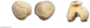
The patella is a sesamoid bone embedded in the quadriceps tendon. It protects the knee joint and improves leverage for the quadriceps muscle.
Base: Superior border for quadriceps tendon attachment.
Apex: Inferior point for patellar ligament attachment.
Lateral and Medial Facets: Articulate with femoral condyles.
Articular Surface: Smooth area for joint movement.
Tibia and Fibula: Bones of the Leg
Structure and Bone Markings of Tibia and Fibula
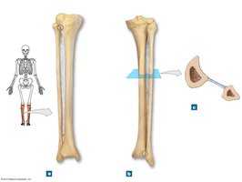
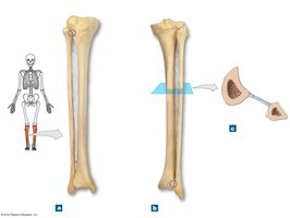
The tibia and fibula are the two bones of the lower leg. The tibia is the main weight-bearing bone, while the fibula provides lateral stability.
Tibia: Medial bone, supports body weight.
Fibula: Lateral bone, stabilizes the ankle.
Intercondylar tubercles: Located between tibial condyles.
Tibial tuberosity: Attachment for patellar ligament.
Medial and Lateral Malleolus: Bony prominences at the ankle.
Ankle and Foot Bones
Structure of the Ankle and Foot
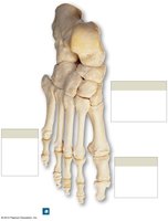
The ankle and foot consist of several bones that provide support, balance, and mobility. The arrangement of these bones forms arches that absorb shock and facilitate movement.
Calcaneus: Heel bone, largest tarsal bone.
Talus: Articulates with tibia and fibula.
Navicular, Cuboid, Cuneiform Bones: Contribute to the structure of the foot.
Metatarsals: Five long bones of the foot.
Phalanges: Toe bones.
Arches: Longitudinal and transverse arches provide strength and flexibility.
Summary Table: Major Lower Appendage Bones and Features
Bone | Main Features | Function |
|---|---|---|
Os Coxa (Hip Bone) | Ilium, Ischium, Pubis, Acetabulum | Supports trunk, attaches lower limb |
Femur | Head, Neck, Trochanters, Condyles | Thigh movement, weight bearing |
Patella | Base, Apex, Facets | Protects knee, improves leverage |
Tibia | Tuberosity, Malleolus | Main weight-bearing leg bone |
Fibula | Head, Malleolus | Stabilizes ankle |
Foot Bones | Calcaneus, Talus, Metatarsals, Phalanges | Support, balance, movement |
Key Terms and Definitions
Acetabulum: The socket in the hip bone for the femur.
Trochanter: Large bony projection for muscle attachment.
Malleolus: Bony prominence at the ankle.
Pelvic brim: The edge separating the false and true pelvis.
Phalanges: Bones of the toes.
Example: Clinical Relevance
Fractures of the femur or pelvis can significantly impact mobility and require surgical intervention. The structure of the pelvis is crucial in obstetrics for childbirth.
Additional info: Academic context was added to clarify bone functions, clinical relevance, and anatomical terminology.
