 Back
BackStudy Guide: Muscular System (Chapter 9)
Study Guide - Smart Notes
 Tailored notes based on your materials, expanded with key definitions, examples, and context.
Muscular System Overview
Introduction to the Muscular System
The muscular system is composed of skeletal, smooth, and cardiac muscles, each playing a vital role in movement, posture, heat production, and glycemic control. Muscles are attached to bones via tendons, enabling voluntary movement, while smooth and cardiac muscles regulate involuntary actions such as passage of substances and control of body openings.
Skeletal muscle: Responsible for voluntary movements, posture, and heat production.
Smooth muscle: Controls involuntary movements in organs and passageways.
Cardiac muscle: Specialized for continuous rhythmic contraction of the heart.
Histology of Muscle Tissue
Types of Muscle Fibers
Muscle tissue can be classified into three types based on microscopic structure and function.
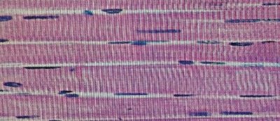
Skeletal Muscle Fiber: Long, cylindrical, multinucleated fibers with visible striations. Responsible for voluntary movement.
Smooth Muscle Fiber: Spindle-shaped, single nucleus, no striations. Found in walls of hollow organs.
Cardiac Muscle Fiber: Branched, usually one or two nuclei, striated, contains intercalated discs. Found only in the heart.
Structure of Intercalated Discs
Intercalated discs are specialized junctions between cardiac muscle cells that allow rapid transmission of electrical impulses and mechanical connection.
Function: Synchronize contraction of cardiac muscle fibers.
Location: Only found in cardiac muscle due to its need for coordinated contraction.
Skeletal Muscle Fiber Anatomy
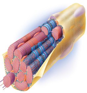
Microscopic Anatomy of Skeletal Muscle
Skeletal muscle fibers are organized into bundles and contain specialized structures for contraction.
Endomysium: Connective tissue surrounding each muscle fiber.
Sarcolemma: Plasma membrane of the muscle cell.
Sarcoplasm: Cytoplasm of the muscle cell.
Sarcoplasmic reticulum: Stores calcium ions necessary for contraction.
Terminal cisternae: Enlarged areas of the sarcoplasmic reticulum adjacent to T tubules.
T tubule: Invaginations of the sarcolemma that help transmit action potentials.
Triad: Structure formed by a T tubule and two terminal cisternae.
Myofibril: Rod-like units within muscle fibers containing contractile proteins.
Nucleus: Multiple nuclei per fiber, located peripherally.
Mitochondria: Provide energy for contraction.
Sarcomere: Functional unit of muscle contraction.
Myofilaments: Actin (thin) and myosin (thick) filaments responsible for contraction.
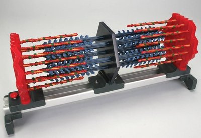
Sarcomere Structure
The sarcomere is the basic contractile unit of skeletal muscle, defined by the arrangement of myofilaments.
A band: Dark region containing thick (myosin) filaments.
I band: Light region containing thin (actin) filaments.
Z disc: Boundary of the sarcomere, anchors thin filaments.
M line: Center of the sarcomere, anchors thick filaments.
Thick filaments: Composed of myosin heads and tails.
Thin filaments: Composed of actin, tropomyosin, and troponin.
Neuromuscular Junction
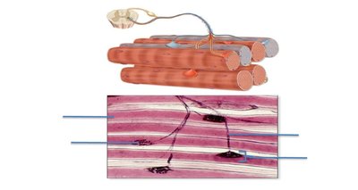
Structure and Function
The neuromuscular junction is the site where a motor neuron communicates with a skeletal muscle fiber to initiate contraction.
Axon branches: Terminal branches of the motor neuron.
Axon terminal: End of the neuron, releases neurotransmitter.
Neuromuscular junction: Synapse between neuron and muscle fiber.
Skeletal muscle fiber: Receives signal to contract.
Skeletal Muscles and Their Actions
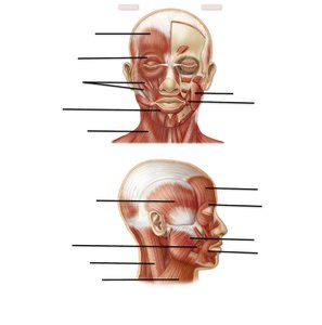
Muscles of the Head and Neck
Muscles in the head and neck region control facial expressions, mastication, and movement of the head.
Frontalis: Raises eyebrows.
Orbicularis oculi: Closes eye, blinking, squinting.
Zygomaticus: Smiling.
Orbicularis oris: Closes mouth, purses lips.
Masseter: Elevates mandible, chewing.
Temporalis: Elevates mandible.
Buccinator: Suckling.
Platysma: Depresses mandible.
Sternocleidomastoid: Flexes and rotates head.
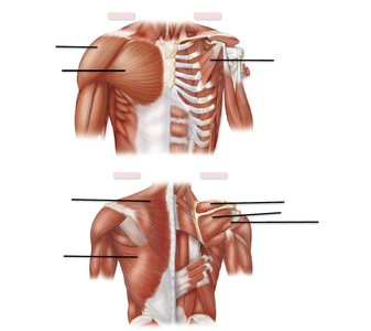
Muscles of the Shoulder and Arm
These muscles facilitate movement of the shoulder and arm, including flexion, extension, abduction, and stabilization.
Pectoralis major: Flexes and adducts arm.
Pectoralis minor: Pulls scapula forward.
Deltoid: Extends and abducts arm.
Trapezius: Elevates scapula.
Latissimus dorsi: Adducts arm.
Rotator cuff muscles (SITS): Stabilize shoulder joint.
Supraspinatus
Infraspinatus
Teres minor
Subscapularis
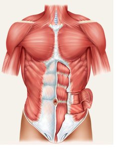
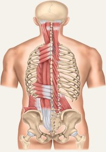
Abdominal and Vertebral Muscles
These muscles support the trunk, compress the abdomen, and allow flexion and extension of the spine.
Rectus abdominis: Flexes spine.
External oblique/aponeurosis: Compresses abdomen.
Internal oblique: Compresses abdomen.
Transversus abdominis: Compresses abdomen.
Erector spinae: Extends spine.
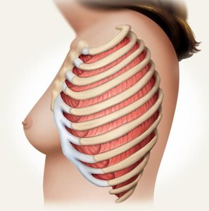
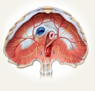
Respiratory Muscles
Muscles involved in breathing include the intercostals and diaphragm.
External intercostals: Inspiration.
Internal intercostals: Expiration.
Diaphragm: Inspiration.
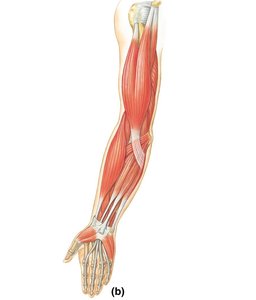
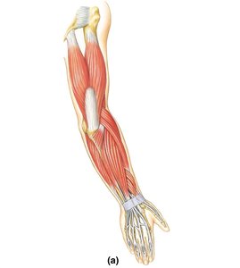
Muscles of the Upper Limb
Muscles of the upper limb are responsible for flexion and extension of the forearm.
Biceps: Flexes forearm.
Brachialis: Flexes forearm.
Brachioradialis: Flexes forearm.
Triceps: Extends forearm.
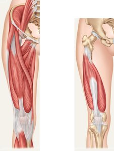
Muscles of the Lower Limb
Anterior View:
Iliopsoas: Flexes thigh.
Tensor fasciae latae: Flexes thigh.
Gracilis: Adducts thigh.
Quadriceps femoris: Extends knee.
Rectus femoris
Vastus lateralis
Vastus medialis
Vastus intermedius
Sartorius: Flexes thigh, medially rotates leg.
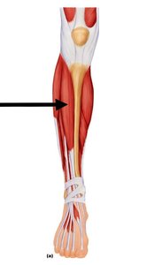
Tibialis anterior: Dorsiflexion of foot.
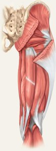
 Posterior View:
Gluteus maximus: Extends and laterally rotates thigh.
Gluteus medius: Abducts hip.
Hamstring muscles: Flex knee.
Biceps femoris
Semimembranosus
Semitendinosus
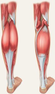
Gastrocnemius (Calcaneal tendon): Plantar flexion of foot.
Soleus (Calcaneal tendon): Plantar flexion of foot.
Check Your Understanding
Review Questions
Name two muscles in the body that have straight muscle fibers: Rectus abdominis, Sartorius.
Name the muscle that has an antagonistic action to: Triceps brachii: Biceps brachii Rectus femoris: Hamstring group
When turning the head to the right side, which sternocleidomastoid is contracting? Left sternocleidomastoid.
Which two muscles synergistically close the jaw? Masseter, Temporalis.
Which is the deepest abdominal muscle? Transversus abdominis.
Which muscles are responsible for inspiration? External intercostals, Diaphragm.
Due to nerve damage, a patient has a clinical condition called drop foot. His toes are dragging and he has difficulty lifting them while walking. Which muscle is affected? Tibialis anterior.
Which muscles will extend the arms during pushups? Triceps brachii.
Winking, blinking and squinting are muscular actions of Orbicularis oculi.
Baseball pitchers are more prone to injury of the rotator cuff group of muscles.
Additional info:
The muscular system is essential for voluntary and involuntary movements, and its structure is closely related to function.
Muscle contraction is regulated by the interaction of actin and myosin within the sarcomere, and is initiated by signals at the neuromuscular junction.
Understanding muscle anatomy and function is critical for diagnosing and treating musculoskeletal disorders.
