 Back
Study Guide: Muscular System – Structure, Classification, and Major Muscles
Study Guide - Smart Notes
 Tailored notes based on your materials, expanded with key definitions, examples, and context.
Muscle Structure and Attachments
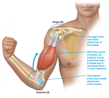
Origin and Insertion
The origin and insertion are fundamental concepts in muscle anatomy. Muscles attach to bones at two points: the origin (the more fixed attachment) and the insertion (the more mobile attachment). During contraction, the insertion moves toward the origin, resulting in movement at the joint the muscle crosses.
Origin: The stable, proximal attachment of the muscle.
Insertion: The movable, distal attachment of the muscle.
Muscle Action: Muscles can only pull, not push.
Joint Crossing: All muscles cross at least one joint, and the bulk of the muscle lies proximal to the joint crossed.
Classification of Muscles
Muscle Roles in Movement
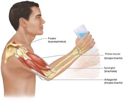
Muscles are classified based on their roles during movement:
Prime Mover (Agonist): The main muscle responsible for a specific movement.
Antagonist: Muscle that opposes the action of the prime mover.
Synergist: Muscle that assists the prime mover.
Fixator: Muscle that stabilizes the origin of the prime mover.
Criteria Used in Naming Muscles
Muscle Naming Conventions
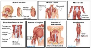
Muscles are named according to several criteria, which help identify their location, shape, function, and other characteristics:
Location: Indicates where the muscle is found (e.g., temporalis, intercostal).
Shape: Describes the muscle's form (e.g., trapezius, rhomboid).
Size: Relative size (e.g., gluteus maximus).
Direction of Fibers: Orientation of muscle fibers (e.g., rectus abdominis).
Number of Origins: Number of tendinous attachments (e.g., biceps brachii).
Location of Attachments: Points of origin and insertion (e.g., sternocleidomastoid).
Action: The movement produced (e.g., flexor digitorum).
Muscles of Facial Expression
Anterior and Lateral Views
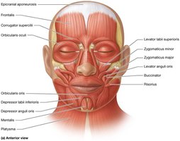
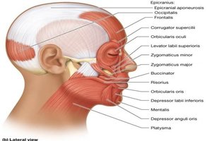
Facial muscles are responsible for expressions and movements such as smiling, frowning, and blinking. They are unique because they insert into the skin rather than bone.
Epicranius (frontalis and occipitalis): Raises eyebrows.
Orbicularis oculi: Closes eyelids.
Orbicularis oris: Closes and protrudes lips.
Zygomaticus major and minor: Elevate corners of the mouth (smiling).
Buccinator: Compresses cheek.
Risorius: Draws corner of mouth laterally.
Platysma: Tenses skin of neck.
Muscles of Mastication
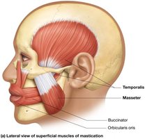
Muscles involved in chewing include:
Temporalis: Elevates and retracts mandible.
Masseter: Elevates mandible.
Buccinator: Assists in mastication by keeping food between teeth.
Orbicularis oris: Closes lips.
Muscles of the Head and Neck
Muscles Involved in Swallowing and Head Movement
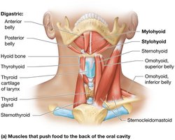
These muscles facilitate swallowing and movement of the head and neck:
Digastric: Elevates hyoid and depresses mandible.
Mylohyoid: Elevates hyoid and floor of mouth.
Stylohyoid: Elevates and retracts hyoid.
Sternohyoid: Depresses hyoid.
Sternocleidomastoid: Flexes and rotates head.
Major Muscles Involved in Common Actions
Facial and Neck Movements
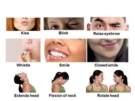
Specific muscles are responsible for distinct actions such as kissing, whistling, smiling, and head movements.
Kiss: Orbicularis oris.
Whistle: Buccinator.
Closed smile: Zygomaticus major.
Extends head: Sternocleidomastoid.
Flexion of neck: Sternocleidomastoid.
Rotate head: Sternocleidomastoid and other neck muscles.
Muscles of the Trunk
Abdominal Muscles
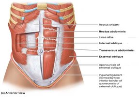
The abdominal muscles support the trunk, allow movement, and hold organs in place by regulating internal pressure.
Rectus abdominis: Flexes trunk, compresses abdominal cavity.
External oblique: Flexes and laterally flexes trunk.
Internal oblique: Flexes and laterally flexes trunk.
Transversus abdominis: Compresses abdominal cavity.
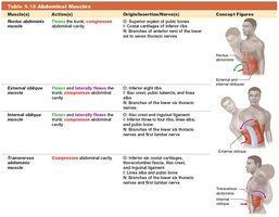
Muscle | Action | Origin/Insertion/Nerves |
|---|---|---|
Rectus abdominis | Flexes the trunk, compresses abdominal cavity | O: Superior aspect of pubic bones I: Costal cartilage of inferior ribs |
External oblique | Flexes and laterally flexes the trunk | O: Inferior eight ribs I: Iliac crest, pubic tubercle, linea alba |
Internal oblique | Flexes and laterally flexes the trunk | O: Iliac crest, inguinal ligament I: Inferior three or four ribs, linea alba, pubic crest |
Transversus abdominis | Compresses abdominal cavity | O: Inferior six costal cartilages, thoracolumbar fascia, iliac crest, inguinal ligament I: Linea alba, pubic crest |
Muscles of the Shoulder and Arm
Superficial and Deep Muscles
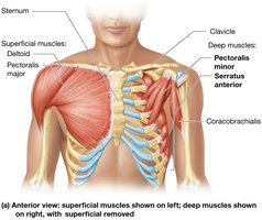
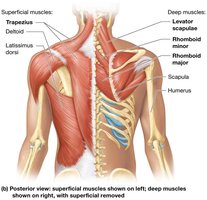
The shoulder and arm muscles are responsible for movements of the upper limb, including flexion, extension, abduction, and rotation.
Deltoid: Abducts arm.
Pectoralis major: Flexes, adducts, and medially rotates arm.
Pectoralis minor: Stabilizes scapula.
Serratus anterior: Protracts scapula.
Coracobrachialis: Flexes and adducts arm.
Trapezius: Elevates, retracts, and rotates scapula.
Latissimus dorsi: Extends, adducts, and medially rotates arm.
Rhomboid major/minor: Retract scapula.
Levator scapulae: Elevates scapula.
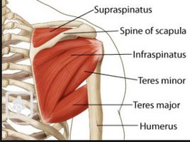
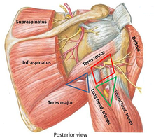
Teres Minor vs. Teres Major
The teres minor and teres major are two distinct muscles of the shoulder:
Teres minor: Rotates arm laterally; part of the rotator cuff.
Teres major: Adducts and medially rotates arm; not part of the rotator cuff.
Muscles of the Arm, Forearm, and Hand
Anterior and Posterior Views
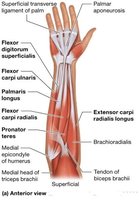
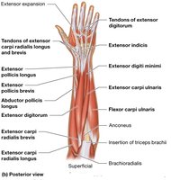
These muscles control movements of the elbow, wrist, and fingers:
Biceps brachii: Flexes elbow, supinates forearm.
Triceps brachii: Extends elbow.
Brachialis: Flexes elbow.
Flexor digitorum superficialis: Flexes fingers.
Extensor carpi radialis longus: Extends and abducts wrist.
Palmaris longus: Flexes wrist.
Pronator teres: Pronates forearm.
Superficial Muscles: Anterior and Posterior Views
Overview of Major Muscles
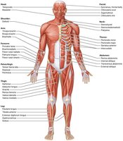
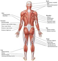
Superficial muscles are those closest to the skin and are responsible for the visible contours of the body. They include muscles of the chest, abdomen, arms, legs, and back.
Anterior view: Shows muscles such as pectoralis major, rectus abdominis, quadriceps, and biceps brachii.
Posterior view: Shows muscles such as trapezius, latissimus dorsi, gluteus maximus, hamstrings, and triceps brachii.
Muscles that Move the Thigh and Knee
Superficial and Deep Muscles
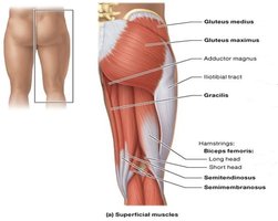
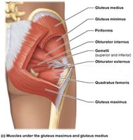
These muscles are essential for walking, running, and maintaining posture:
Gluteus maximus: Extends and laterally rotates thigh.
Gluteus medius: Abducts and medially rotates thigh.
Adductor magnus: Adducts thigh.
Hamstrings (biceps femoris, semitendinosus, semimembranosus): Flex knee, extend thigh.
Gracilis: Adducts thigh, flexes knee.
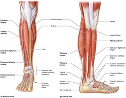
Muscles That Move the Ankle and Foot
Anterior and Lateral Views
Muscles of the lower leg control movements of the ankle and foot, including dorsiflexion, plantarflexion, inversion, and eversion.
Tibialis anterior: Dorsiflexes and inverts foot.
Fibularis longus: Everts and plantarflexes foot.
Extensor digitorum longus: Extends toes, dorsiflexes foot.
Gastrocnemius: Plantarflexes foot, flexes knee.
Soleus: Plantarflexes foot.
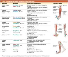
Muscle | Action | Origin/Insertion/Nerves |
|---|---|---|
Tibialis anterior | Dorsiflexes the foot | O: Lateral condyle and proximal diaphysis of tibia I: Medial cuneiform and first metatarsal |
Fibularis longus | Everts and plantarflexes the foot | O: Head and proximal fibula I: Medial cuneiform and first metatarsal |
Extensor digitorum longus | Extends toes, dorsiflexes foot | O: Lateral condyle of tibia, proximal fibula I: Phalanges of toes |
Gastrocnemius | Plantarflexes foot, flexes knee | O: Medial and lateral condyles of femur I: Calcaneus via Achilles tendon |
Soleus | Plantarflexes foot | O: Head of fibula, medial border of tibia I: Calcaneus via Achilles tendon |
Putting It All Together: The Big Picture of Muscle Movement
Integrated Muscle Actions
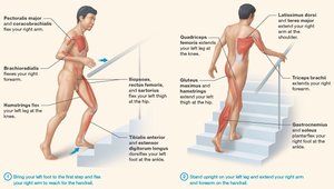
Muscle movement is a coordinated process involving multiple muscle groups. For example, walking up stairs requires the activation of muscles in the chest, arms, thighs, and lower legs to produce smooth, efficient movement.
Pectoralis major and coracobrachialis: Flex arm.
Brachialis: Flexes elbow.
Hamstrings: Flex knee.
Quadriceps: Extend knee.
Gluteus maximus: Extends thigh.
Triceps brachii: Extends elbow.
Tibialis anterior: Dorsiflexes foot.
Gastrocnemius: Plantarflexes foot.
Summary Table: Muscle Classification and Naming Criteria
Comparison of Muscle Naming Criteria
Criterion | Example | Description |
|---|---|---|
Location | Temporalis | Located near the temporal bone |
Shape | Trapezius | Trapezoid-shaped muscle |
Size | Gluteus maximus | Largest gluteal muscle |
Direction of fibers | Rectus abdominis | Fibers run straight |
Number of origins | Biceps brachii | Two origins |
Location of attachments | Sternocleidomastoid | Attaches to sternum, clavicle, and mastoid process |
Action | Flexor digitorum | Flexes fingers |
Key Equations and Concepts
Muscle Contraction Principle
Muscle contraction results in the insertion moving toward the origin, producing movement at the joint.
Force Generation
The force generated by a muscle is proportional to its cross-sectional area and the number of muscle fibers activated.
Equation for muscle force:
Where F is force, A is cross-sectional area, and σ is specific tension.
Conclusion
The muscular system is essential for movement, posture, and various bodily functions. Understanding muscle structure, classification, and naming conventions provides a foundation for further study in anatomy and physiology.
