 Back
Chapter 12
Study Guide - Smart Notes
 Tailored notes based on your materials, expanded with key definitions, examples, and context.
Q1. Match the anatomical and functional divisions of the nervous system with their descriptions.
Background
Topic: Organization of the Nervous System
This question tests your understanding of how the nervous system is divided both anatomically (CNS vs. PNS) and functionally (sensory/afferent vs. motor/efferent, somatic vs. autonomic).
Key Terms:
CNS (Central Nervous System): Brain and spinal cord.
PNS (Peripheral Nervous System): Nerves outside the CNS.
Afferent Division: Sensory input to CNS.
Efferent Division: Motor output from CNS.
Somatic Nervous System: Voluntary control of skeletal muscles.
Autonomic Nervous System: Involuntary control of smooth/cardiac muscle and glands.
Step-by-Step Guidance
Read each definition carefully and identify keywords (e.g., 'sensory', 'motor', 'skeletal muscle', 'smooth muscle').
Recall that the afferent division is also called the sensory division and carries information to the CNS.
The efferent division is also called the motor division and carries commands from the CNS to effectors (muscles/glands).
Distinguish between the somatic (voluntary, skeletal muscle) and autonomic (involuntary, smooth/cardiac muscle) systems.
Match the CNS and PNS with their anatomical components (brain/spinal cord vs. nerves).
Try solving on your own before revealing the answer!
Q2. Complete the table of functional divisions of the peripheral nervous system (PNS) using the terms: Autonomic, Motor, Sensory, Somatic.
Background
Topic: Functional Organization of the PNS
This question checks your ability to classify the PNS into its voluntary (somatic) and involuntary (autonomic) branches, and to distinguish between sensory and motor functions.
Key Terms:
Sensory (Afferent): Carries information to CNS.
Motor (Efferent): Carries commands from CNS.
Somatic: Voluntary, skeletal muscle.
Autonomic: Involuntary, smooth/cardiac muscle and glands.
Step-by-Step Guidance
Draw a table with two main branches under PNS: Sensory and Motor.
Under Motor, split into Somatic (voluntary) and Autonomic (involuntary).
Place the terms in the correct spots based on their definitions.
Try solving on your own before revealing the answer!
Q3. Indicate whether each structure is part of the peripheral (P) or central (C) nervous system.
Background
Topic: Anatomical Divisions of the Nervous System
This question tests your ability to distinguish between structures found in the CNS (brain, spinal cord, tracts) and PNS (nerves).
Key Terms:
Brain, Spinal Cord, Tract: CNS
Nerve: PNS
Step-by-Step Guidance
Recall that the CNS includes the brain and spinal cord, and tracts are bundles of axons within the CNS.
The PNS includes nerves (bundles of axons outside the CNS).
Assign 'C' or 'P' to each term based on its location.
Try solving on your own before revealing the answer!
Q4. Label the following image of a neuron. Add a myelin sheath to the image.
Background
Topic: Neuron Structure
This question tests your ability to identify the main parts of a neuron and understand the role of myelin in nerve conduction.
Key Terms:
Dendrites: Receive signals.
Cell body (soma): Contains nucleus and organelles.
Axon hillock: Where action potentials begin.
Axon: Conducts impulses away from cell body.
Myelin: Insulating layer around axon.
Nodes of Ranvier: Gaps in myelin sheath.
Axon terminals: Release neurotransmitters.
Postsynaptic neuron: Receives signal.
Step-by-Step Guidance
Identify the cell body (soma) at the center of the neuron.
Locate the dendrites branching from the soma.
Find the axon hillock at the base of the soma, where the axon begins.
Trace the axon away from the soma, noting the myelin sheath (insulating segments) and nodes of Ranvier (gaps).
Label the axon terminals at the end of the axon, and indicate the postsynaptic neuron where the signal is sent.
Try labeling the image and adding the myelin sheath before checking the answer!
Q5. Explain the gray and white matter of the spinal cord and the brain.
Background
Topic: CNS Structure
This question tests your understanding of the composition and function of gray and white matter in the CNS.
Key Terms:
Gray matter: Contains neuron cell bodies, dendrites, and unmyelinated axons.
White matter: Contains myelinated axons.
Step-by-Step Guidance
Describe where gray and white matter are located in the brain and spinal cord (e.g., gray matter is outer in brain, inner in spinal cord).
Explain the functional significance of each (processing vs. transmission of signals).
Relate the color difference to the presence or absence of myelin.
Try explaining in your own words before revealing the answer!
Q6. What is the purpose of gated sodium and potassium channels in the membrane of a neuron?
Background
Topic: Neurophysiology – Ion Channels
This question tests your understanding of how neurons generate and propagate electrical signals using ion channels.
Key Terms:
Gated channels: Open or close in response to stimuli.
Sodium (Na+) channels: Involved in depolarization.
Potassium (K+) channels: Involved in repolarization.
Step-by-Step Guidance
Explain how opening sodium channels allows Na+ to enter the neuron, causing depolarization.
Describe how opening potassium channels allows K+ to exit, causing repolarization.
Relate these changes to the generation and propagation of action potentials.
Try describing the purpose of these channels before revealing the answer!
Q7. Match the parts of a multipolar neuron with their functions.
Background
Topic: Neuron Anatomy and Function
This question tests your ability to identify neuron structures and their roles in signal transmission.
Key Terms:
Review the definitions for each part (axon, myelin, dendrites, etc.) and their functions (signal transmission, insulation, neurotransmitter release, etc.).
Step-by-Step Guidance
Read each function and match it to the correct neuron part based on your knowledge of neuron structure.
Pay attention to terms like 'soma', 'axon hillock', 'Nissl bodies', and 'synaptic cleft'.
Use process of elimination for any terms you are unsure about.
Try matching each part before revealing the answer!
Q8. Match the types of neuroglia with their descriptions.
Background
Topic: Neuroglia (Glial Cells)
This question tests your knowledge of the different types of glial cells in the CNS and PNS and their functions.
Key Terms:
Astrocytes: Blood-brain barrier, support.
Oligodendrocytes: Myelinate CNS axons.
Ependymal cells: Line ventricles, produce CSF.
Schwann cells: Myelinate PNS axons.
Microglia: Immune defense in CNS.
Step-by-Step Guidance
Review the unique functions and locations of each glial cell type.
Match each description to the correct cell based on keywords (e.g., 'myelin in PNS' = Schwann cell).
Remember that some cells are only found in the CNS or PNS.
Try matching each neuroglia before revealing the answer!
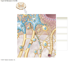
Q9. Label the types of neuroglia in the CNS in the image.
Background
Topic: CNS Neuroglia Identification
This question tests your ability to visually identify astrocytes, oligodendrocytes, microglia, and ependymal cells in CNS tissue.
Key Terms:
Astrocytes: Star-shaped, support neurons.
Oligodendrocytes: Myelinate CNS axons.
Microglia: Small, immune cells.
Ependymal cells: Line ventricles, produce CSF.
Step-by-Step Guidance
Look for the star-shaped cells (astrocytes), cells wrapping axons (oligodendrocytes), small round cells (microglia), and cells lining cavities (ependymal cells).
Label each cell type in the image based on their appearance and location.
Try labeling the image before revealing the answer!
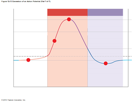
Q17. Label the graph of an action potential using the key provided.
Background
Topic: Action Potential Phases
This question tests your understanding of the sequence of events during an action potential, including depolarization, repolarization, and the closing of ion channels.
Key Terms:
Resting membrane potential: Baseline voltage.
Depolarization: Na+ enters, membrane potential rises.
Repolarization: K+ leaves, membrane potential falls.
Closing of Na+ channels: Marks the end of depolarization.
Potassium channels close: End of repolarization.
Step-by-Step Guidance
Identify the baseline (resting membrane potential) on the graph.
Locate the steep upward slope (depolarization, Na+ enters).
Find the peak where Na+ channels close.
Trace the downward slope (repolarization, K+ leaves) and mark where potassium channels close.
Label each phase according to the key provided.
Try labeling the graph before revealing the answer!
