 Back
Study Guide: Nervous System Organization, Neurons, Neuroglia, and Pathologies
Study Guide - Smart Notes
 Tailored notes based on your materials, expanded with key definitions, examples, and context.
Nervous System Organization
Central and Peripheral Nervous Systems
The nervous system is divided into the Central Nervous System (CNS) and Peripheral Nervous System (PNS). The CNS consists of the brain and spinal cord, while the PNS includes all other neural elements.
CNS: Responsible for processing and interpreting sensory information and issuing motor commands.
PNS: Transmits sensory and motor signals between the CNS and the rest of the body.
Somatic vs. Autonomic Nervous Systems
The PNS is further divided into the somatic and autonomic nervous systems. The somatic system controls voluntary movements, while the autonomic system regulates involuntary functions.
Somatic Nervous System: Controls skeletal muscle contractions.
Autonomic Nervous System: Regulates smooth muscle, cardiac muscle, and glands. Divided into sympathetic (fight or flight) and parasympathetic (rest and digest) branches.
Neurons: Structure and Function
Basic Anatomy of a Neuron
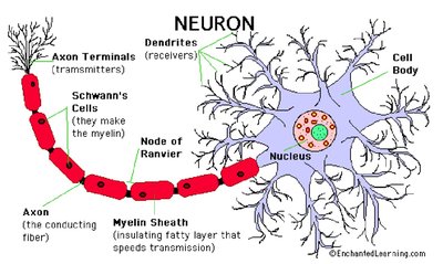
Neurons are specialized cells that transmit electrical and chemical signals. Their structure includes dendrites, a cell body, an axon, and axon terminals.
Dendrites: Receive incoming signals.
Cell Body (Soma): Contains the nucleus and organelles.
Axon: Conducts impulses away from the cell body.
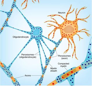
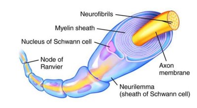
Myelin Sheath: Insulates the axon, speeding up signal transmission.
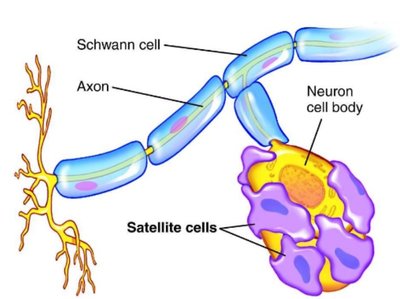
Schwann Cells: Produce myelin in the PNS.
Node of Ranvier: Gaps in myelin sheath that facilitate rapid conduction.
Neuron Types
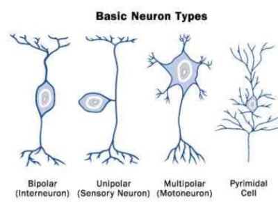
Neurons are classified based on their structure and function.
Bipolar: One axon and one dendrite (found in sensory organs).
Unipolar: Single process that splits into two branches (sensory neurons).
Multipolar: Multiple dendrites and one axon (motor neurons).
Pyramidal: Found in the cerebral cortex.
Synapse and Neurotransmission
Synaptic Transmission
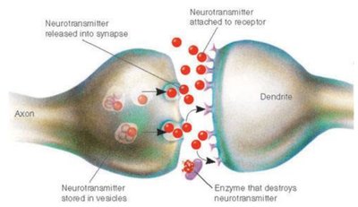
The synapse is the junction between two neurons or a neuron and an effector cell. Neurotransmitters are released from the axon terminal, cross the synaptic cleft, and bind to receptors on the postsynaptic cell.
Enzyme degradation: Enzymes break down neurotransmitters (e.g., acetylcholinesterase for acetylcholine).
Diffusion: Neurotransmitters diffuse away from the synapse.
Reuptake: Neurotransmitters are reabsorbed by the presynaptic neuron.
Synapse Types
Synapses are classified by their location:
Axodendritic: Axon to dendrite.
Axosomatic: Axon to soma (cell body).
Axoaxonic: Axon to axon.
Neuroglia (Glial Cells)
Types and Functions
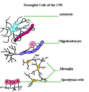
Neuroglia are support cells for neurons, found in both CNS and PNS. They undergo mitosis and perform various functions.
Microglia: Phagocytic cells that remove debris and attack invaders.
Oligodendrocytes: Produce myelin sheath in CNS.
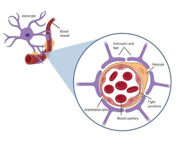
Astrocytes: Support neurons, maintain electrolyte balance, and form part of the blood-brain barrier.
Ependymal Cells: Produce cerebrospinal fluid (CSF).
Schwann Cells: Produce myelin in PNS.
Satellite Cells: Support ganglia in PNS.
Pathologies of the Nervous System
Multiple Sclerosis (MS)
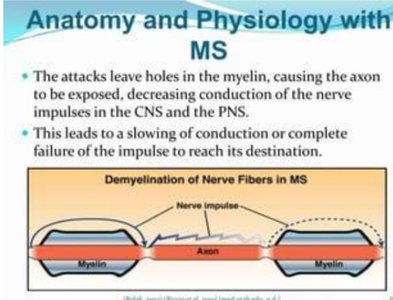
MS is an autoimmune disorder where the immune system attacks the myelin sheath, leading to impaired nerve conduction and formation of plaques.
Symptoms: Slowed or failed nerve impulse transmission.
Pathology: Demyelination in CNS and PNS.
Tetanus and Botulism
Tetanus: Caused by Clostridium tetani toxin, inhibits release of GABA and glycine, resulting in spastic paralysis.
Botulism: Inhibits acetylcholine release, causing flaccid paralysis.
Neurotransmitter Removal and Clinical Relevance
Serotonin: Removed by reuptake; SSRIs block reuptake to treat depression.
Acetylcholine: Removed by acetylcholinesterase.
Spinal Cord Structure and Function
Gray and White Matter
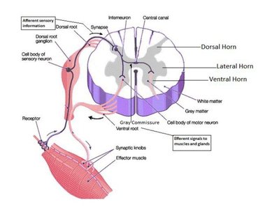
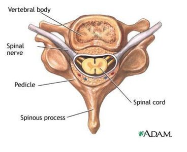
Gray Matter: Contains cell bodies, no myelin.
White Matter: Contains myelinated axons.
Spinal Cord Anatomy
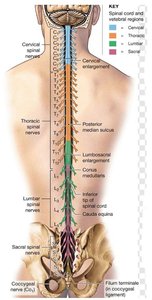
Conus Medullaris: End of solid spinal cord at L1.
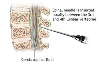
Cauda Equina: Bundle of nerve fibers below L1.
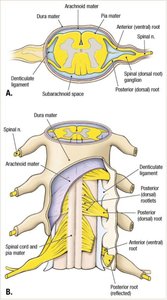
Denticulate Ligaments: Anchor spinal cord to meninges.
Filum Terminale: Extension of pia mater anchoring cord to coccyx.
Spinal Cord Pathologies
Shingles and Poliovirus
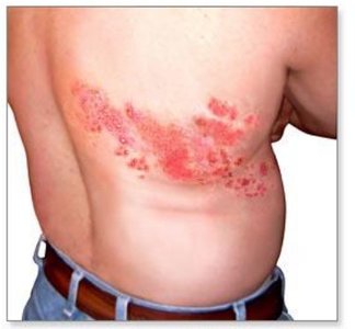
Shingles: Reactivation of Varicella Zoster Virus in dorsal root ganglion.
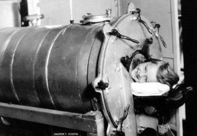
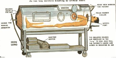
Poliovirus: Attacks anterior horn cells, may cause respiratory failure.
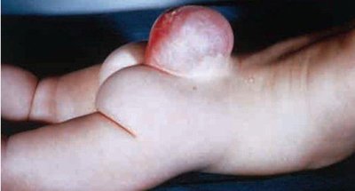
Spina Bifida
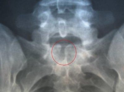
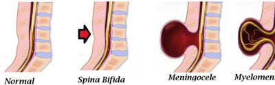
Spina bifida is a congenital defect where the vertebral column does not close properly.
Spina Bifida Occulta: Malfusion of sacrum, no damage to cauda equina.
Meningocele: Meninges protrude through vertebral defect.
Myelomeningocele: Both meninges and spinal cord protrude.
Brain Structure and Function
Gray and White Matter in the Brain
Cortex: Outer gray matter (cell bodies).
White Matter: Inner axons.
Gyri: Ridges.
Sulci: Depressions.
Cerebrum and Cerebellum
Cerebrum: Responsible for thinking, reasoning, feeling, interpreting, personality, and behavior.
Cerebellum: Balance, muscle coordination, timekeeping, and sensory evaluation.
Corpus Callosum
Corpus Callosum: White matter connecting left and right hemispheres.
Thalamus and Hypothalamus
Thalamus: Relay center for sensory information.
Hypothalamus: Controls hormones, appetite, thirst, temperature, sex drive.
Pituitary Gland
Pituitary: Controlled by hypothalamus, rests in sella turcica.
Brain Stem
Midbrain: Connects pons to rest of brain.
Pons: Relay centers, cranial nerve functions.
Medulla Oblongata: Controls heart rate, respiration, reflexes.
Cerebrospinal Fluid (CSF) and Meninges
CSF Production and Flow
CSF is produced by the choroid plexus in the ventricles and flows through the brain and spinal cord.
Choroid Plexus: Lined with ependymal cells, forms blood-CSF barrier.
CSF Pathway: Lateral ventricles → Foramen of Monro → Third ventricle → Cerebral Aqueduct → Fourth ventricle → Median and lateral apertures → Central canal and subarachnoid space.
Hydrocephalus: Caused by improper CSF flow.
Meninges
The meninges are three connective tissue layers protecting the CNS.
Pia Mater: Innermost, delicate.
Arachnoid Mater: Middle, web-like; subarachnoid space contains CSF.
Dura Mater: Outermost, tough; subdural and epidural spaces.
Cranial Nerves Overview
Major Cranial Nerves and Functions
III Oculomotor: Eye movement, iris, accommodation.
IV Trochlear: Superior oblique muscle.
V Trigeminal: Mastication, facial sensation.
VI Abducens: Lateral rectus muscle.
VII Facial: Facial expression, taste, glands.
VIII Vestibulocochlear: Hearing, balance.
IX Glossopharyngeal: Pharyngeal muscles, taste, blood pressure.
X Vagus: Swallowing, thoracic/abdominal organs.
XI Accessory: Swallowing, neck muscles.
XII Hypoglossal: Tongue muscles.
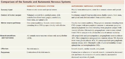
Summary Table: Somatic vs. Autonomic Nervous Systems
Feature | Somatic Nervous System | Autonomic Nervous System |
|---|---|---|
Sensory Input | From somatic senses and special senses | Mainly from interoceptors, some from somatic and special senses |
Control of Motor Output | Voluntary control from cerebral cortex | Involuntary control from hypothalamus, limbic system, brain stem, spinal cord |
Motor Neuron Pathway | One-neuron pathway | Usually two-neuron pathway |
Neurotransmitters | Acetylcholine (ACh) | ACh, norepinephrine (NE), epinephrine |
Effectors | Skeletal muscle | Cardiac muscle, smooth muscle, glands |
Responses | Contraction of skeletal muscle | Contraction/relaxation of smooth muscle, increased/decreased gland secretion |
