 Back
Study Guide: Nervous System Structure and Function (A&P College Level)
Study Guide - Smart Notes
 Tailored notes based on your materials, expanded with key definitions, examples, and context.
Nervous System Overview
Central and Peripheral Nervous Systems
The nervous system is divided into the Central Nervous System (CNS) and Peripheral Nervous System (PNS). The CNS consists of the brain and spinal cord, while the PNS includes all other neural elements. The PNS is further divided into the autonomic and somatic nervous systems.
CNS: Brain and spinal cord; responsible for processing and integrating information.
PNS: Nerves outside the CNS; transmits sensory and motor signals.
Somatic NS: Controls voluntary movements (skeletal muscles).
Autonomic NS: Controls involuntary functions (heart, glands, smooth muscle).
Neuron Structure and Function
Basic Anatomy of a Neuron
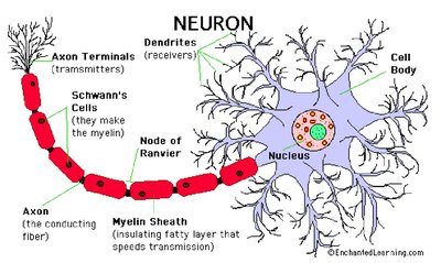
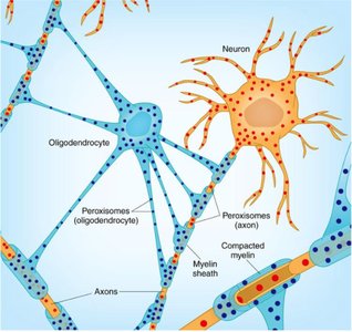
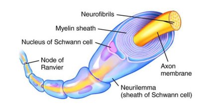
Neurons are specialized cells that transmit electrical and chemical signals. They consist of a cell body, dendrites, axon, and axon terminals. The myelin sheath, produced by glial cells, insulates the axon and speeds up signal transmission.
Dendrites: Receive incoming signals.
Cell Body (Soma): Contains the nucleus and organelles.
Axon: Conducts impulses away from the cell body.
Myelin Sheath: Insulating layer that increases conduction speed.
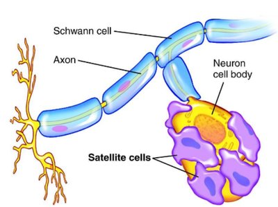
Schwann Cells: Produce myelin in the PNS.
Nodes of Ranvier: Gaps in myelin sheath where action potentials are regenerated.
Types of Neurons
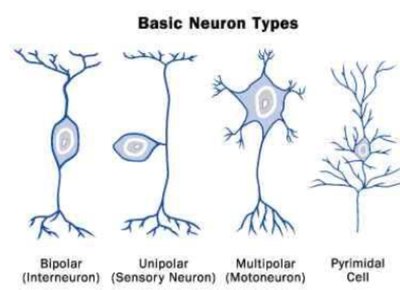
Neurons are classified based on their structure and function:
Bipolar: One axon and one dendrite (found in sensory organs).
Unipolar: Single process that splits into two branches (sensory neurons).
Multipolar: One axon, multiple dendrites (most common, motor neurons).
Pyramidal Cell: Found in the cerebral cortex.
Synapse and Neurotransmission
Synaptic Transmission
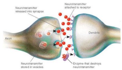
A synapse is the junction between two neurons or a neuron and an effector cell. Neurotransmitters are released from the presynaptic neuron and bind to receptors on the postsynaptic cell, triggering a response.
Neurotransmitter Release: Stored in vesicles, released into synaptic cleft.
Receptor Binding: Neurotransmitters bind to postsynaptic receptors.
Removal: Enzyme degradation, diffusion, or reuptake.
Types of Synapses
Synapses are classified by their location:
Axodendritic: Axon to dendrite.
Axosomatic: Axon to soma (cell body).
Axoaxonic: Axon to axon.
Glial Cells (Neuroglia)
Central Nervous System Glial Cells
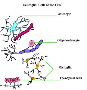
Glial cells support neurons and maintain homeostasis. They are essential for neural function and undergo mitosis.
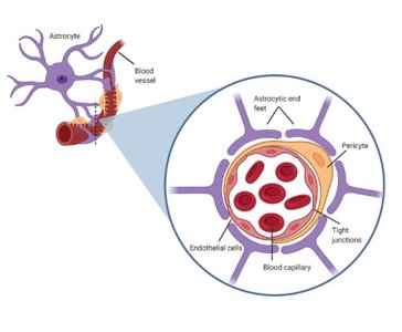
Astrocytes: Support neurons, maintain electrolyte balance, form blood-brain barrier.
Oligodendrocytes: Produce myelin sheath in CNS.
Microglia: Phagocytic cells, remove debris and attack invaders.
Ependymal Cells: Produce cerebrospinal fluid (CSF).
Peripheral Nervous System Glial Cells
Schwann Cells: Produce myelin in the PNS.
Satellite Cells: Support ganglia (clusters of nerve cell bodies).
Diseases and Disorders
Multiple Sclerosis (MS)
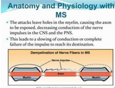
MS is an autoimmune disorder where the immune system attacks the myelin sheath, leading to impaired nerve conduction and formation of plaques (scar tissue).
Symptoms: Slowed or failed nerve impulse transmission.
Pathology: Demyelination in CNS and PNS.
Comparison: Somatic vs. Autonomic Nervous Systems
Functional Differences
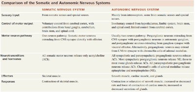
The somatic and autonomic nervous systems differ in their control, effectors, and neurotransmitters.
Somatic NS: Voluntary control, skeletal muscle, acetylcholine as neurotransmitter.
Autonomic NS: Involuntary control, smooth/cardiac muscle and glands, acetylcholine and norepinephrine.
Spinal Cord Structure
Gray and White Matter
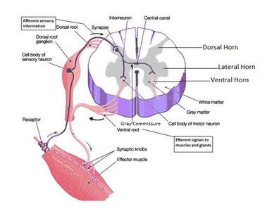
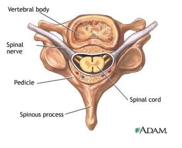
The spinal cord contains gray matter (cell bodies, no myelin) and white matter (myelinated axons). The anterior horn is motor, and the posterior horn is sensory.
Gray Matter: No myelin, contains neuron cell bodies.
White Matter: Myelinated axons.
Summary Table: Somatic vs. Autonomic Nervous Systems
Feature | Somatic Nervous System | Autonomic Nervous System |
|---|---|---|
Sensory Input | From somatic senses and special senses | Mainly from interoceptors, some from somatic and special senses |
Control of Motor Output | Voluntary control from cerebral cortex | Involuntary control from hypothalamus, limbic system, brain stem, spinal cord |
Motor Neuron Pathway | One-neuron pathway | Two-neuron pathway |
Neurotransmitters | Acetylcholine (ACh) | ACh and norepinephrine (NE) |
Effectors | Skeletal muscle | Cardiac muscle, smooth muscle, glands |
Responses | Contraction of skeletal muscle | Contraction/relaxation of muscle, secretion of glands |
