 Back
BackStudy Guide: Sense Organs and Special Senses in Anatomy & Physiology CH 16
Study Guide - Smart Notes
 Tailored notes based on your materials, expanded with key definitions, examples, and context.
Sense Organs: Anatomy & Physiology
Properties and Types of Sensory Receptors
Sensory receptors are specialized structures that detect various stimuli and initiate the process of sensation and perception. They are essential for the nervous system to interpret the environment and internal conditions.
Receptor: A structure specialized to detect a stimulus. Can be a bare nerve ending or a complex sense organ.
Sense Organ: Combines nerve tissue with accessory tissues (epithelial, muscular, connective) to enhance response to specific stimuli.
Transduction: Conversion of stimulus energy (light, heat, touch, sound) into nerve signals.
Sensation: Local electrical change (receptor potential) in response to stimulus; if strong enough, triggers action potentials.
Perception: Conscious interpretation of a stimulus; not all sensations reach perception due to filtering in the CNS.
Four Types of Information Transmitted by Receptors
Modality: Type of stimulus (e.g., vision, hearing, taste); determined by which brain region receives the signal.
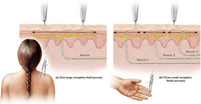
Location: Where the stimulus is detected; encoded by which nerve fibers are firing. Receptive field: Area within which a sensory neuron detects stimuli. Smaller fields allow finer discrimination.
Intensity: Strength of stimulus; encoded by which fibers respond, how many respond, and firing frequency.
Duration: How long the stimulus lasts; encoded by changes in firing frequency. Sensory adaptation: Reduced response to prolonged stimulus.
Phasic receptors: Adapt quickly (e.g., smell, hair movement).
Tonic receptors: Adapt slowly (e.g., body position, muscle tension).
Classification of Receptors
By Modality:
Thermoreceptors: Heat and cold
Photoreceptors: Light (eyes)
Nociceptors: Pain (tissue injury)
Chemoreceptors: Chemicals (odors, tastes, body fluids)
Mechanoreceptors: Physical deformation (touch, pressure, vibration)
By Origin:
Exteroceptors: External stimuli (vision, hearing, touch)
Interoceptors: Internal stimuli (stretch, pressure, visceral pain)
Proprioceptors: Body position and movement (muscles, tendons, joints)
By Distribution:
General senses: Widely distributed (touch, pressure, pain, etc.)
Special senses: Limited to head, complex organs (vision, hearing, equilibrium, taste, smell)
The General Senses
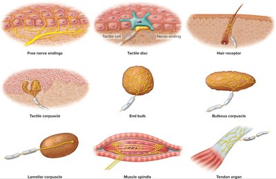
Types of Somatosensory Receptors
Somatosensory receptors are distributed throughout the skin, muscles, and viscera, and are responsible for detecting touch, pressure, temperature, and pain.
Unencapsulated Nerve Endings:
Free nerve endings: Detect temperature and pain
Tactile (Merkel) discs: Detect light touch and texture
Hair receptors: Respond to hair movement
Encapsulated Nerve Endings:
Tactile (Meissner) corpuscles: Light touch and texture
End bulbs: Similar to tactile corpuscles, found in mucous membranes
Bulbous (Ruffini) corpuscles: Heavy touch, pressure, skin stretch
Lamellar (Pacinian) corpuscles: Deep pressure and vibration
Muscle spindles and tendon organs: Proprioception
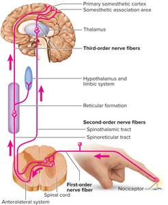
Somatosensory Projection Pathways
Sensory information travels from receptors to the cerebral cortex via a three-neuron pathway.
First-order neuron: From receptor to spinal cord or brainstem
Second-order neuron: Decussates (crosses) and ends in thalamus or cerebellum
Third-order neuron: Thalamus to primary somesthetic cortex
Pain: Types and Mechanisms
Pain is an unpleasant sensation signaling actual or potential tissue damage.
Nociceptive pain: From tissue injury; includes visceral, deep somatic, and superficial somatic pain
Neuropathic pain: From nerve injury
Fast pain: Sharp, localized; carried by myelinated A-delta fibers
Slow pain: Dull, aching; carried by unmyelinated C fibers
Projection Pathways for Pain
Head: Cranial nerves to medulla, thalamus, cortex
Neck and below: Spinothalamic, spinoreticular, and gracile fasciculus tracts
Referred Pain
Pain from internal organs is often perceived as originating from superficial sites due to neural convergence. 
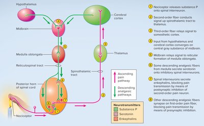
CNS Modulation of Pain
The central nervous system can modulate pain through endogenous opioids and spinal gating mechanisms.
Endogenous opioids: Enkephalins, endorphins, dynorphins block pain and produce pleasure
Spinal gating: Inhibits pain signals at the posterior horn of the spinal cord
Descending analgesic fibers: Arise in brainstem, activate inhibitory interneurons
Rubbing/massaging: Activates mechanoreceptors, inhibits pain transmission
The Chemical Senses
Gustation—The Sense of Taste
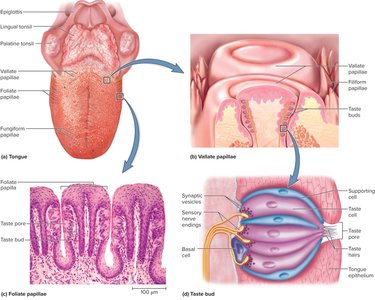
Gustation is the perception of molecules dissolved in water, detected by taste buds mainly on the tongue.
Lingual papillae: Four types—filiform (no taste buds), foliate, fungiform, vallate (contain taste buds)
Taste buds: Clusters of taste cells, supporting cells, and basal cells; taste cells have microvilli (taste hairs) projecting into taste pores
Five primary tastes: Salty, sweet, umami, sour, bitter; possibly oleogustus (fat) and water
Mechanisms:
Sugars, alkaloids, glutamate: Activate G protein-coupled receptors
Sodium, acids: Enter taste cells directly, depolarizing them
Projection pathways: Facial, glossopharyngeal, and vagus nerves carry taste signals to medulla, thalamus, and cortex
Olfaction—The Sense of Smell
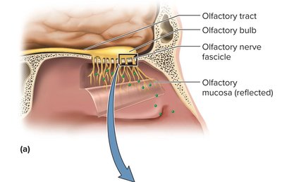
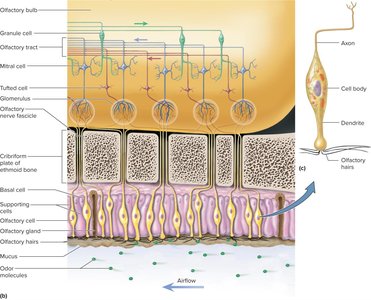
Olfaction is the detection of airborne chemicals (odorants) by olfactory cells in the nasal cavity.
Olfactory mucosa: Contains olfactory cells (neurons), supporting cells, and basal stem cells
Olfactory cell structure: Modified dendrite with olfactory hairs (cilia); axons form olfactory nerve (CN I)
Transduction: Odorant binds G protein-coupled receptor, activates cAMP, opens ion channels, depolarizes membrane
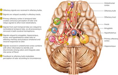
Projection pathways: Olfactory cell axons synapse in olfactory bulbs, then glomeruli, mitral and tufted cells carry signals to primary olfactory cortex, amygdala, hippocampus, insula, hypothalamus
Hearing and Equilibrium
The Nature of Sound
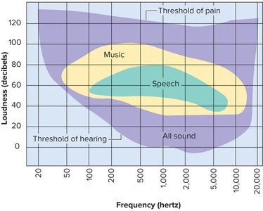
Sound is an audible vibration of molecules, characterized by pitch (frequency) and loudness (amplitude).
Pitch: Determined by frequency (Hz)
Loudness: Measured in decibels (dB)
Humans hear 20–20,000 Hz; normal conversation is 60 dB
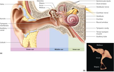
Anatomy of the Ear
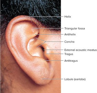
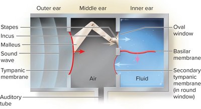
Outer Ear
Auricle (pinna): Funnel for conducting vibrations to eardrum
Auditory canal: Passage to eardrum, protected by hairs and earwax
Middle Ear
Tympanic membrane: Vibrates in response to sound
Tympanic cavity: Air-filled space between outer and inner ear
Auditory ossicles: Malleus, incus, stapes; transmit vibrations to inner ear
Muscles: Stapedius and tensor tympani protect inner ear from loud sounds
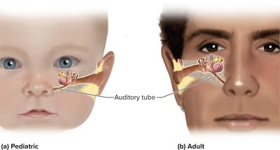
Middle-Ear Infection
Otitis media: Common in children due to short, horizontal auditory tube; can cause hearing loss
Inner Ear
Bony labyrinth: Internal passages in temporal bone
Membranous labyrinth: Fleshy tubes suspended within bony labyrinth
Perilymph: Fluid between labyrinths
Endolymph: Fluid within membranous labyrinth
Vestibule: Contains organs of equilibrium
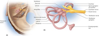
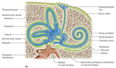
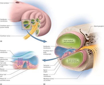
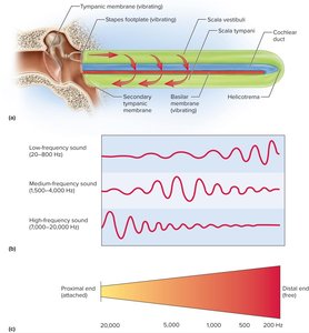
Cochlea
Organ of hearing: Coiled structure with three fluid-filled chambers (scala vestibuli, scala tympani, cochlear duct)
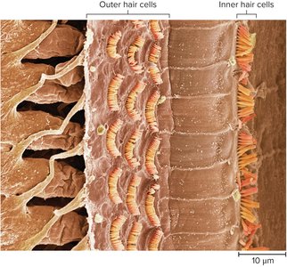
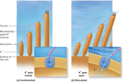
Spiral (acoustic) organ: Converts vibrations to nerve signals; contains hair cells and supporting cells
The Physiology of Hearing
Middle ear: Ossicles concentrate energy, protect inner ear
Stimulation of cochlear hair cells: Vibration causes basilar membrane movement, bending stereocilia, opening K+ channels, depolarizing cells
Sensory Coding
Loudness: Intensity of cochlear vibrations; higher amplitude triggers more action potentials
Pitch: Determined by which part of basilar membrane vibrates (basal end = high pitch, distal end = low pitch)
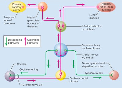
Auditory Projection Pathways
First-order neurons: Spiral ganglion, cochlear nerve
Second-order neurons: Cochlear nucleus, superior olivary nucleus, inferior colliculi
Third-order neurons: Inferior colliculi to thalamus
Fourth-order neurons: Thalamus to primary auditory cortex
Equilibrium
The Physiology of Equilibrium
Vestibular apparatus: Three semicircular ducts and two chambers (saccule, utricle)
Static equilibrium: Orientation of head in space; detected by saccule and utricle
Dynamic equilibrium: Motion or acceleration; linear acceleration detected by saccule and utricle, angular acceleration by semicircular ducts
Macula: Patch of hair cells in saccule and utricle; otolithic membrane weighted with otoliths enhances gravity and motion detection
Semicircular ducts: Detect rotary movements; ampulla contains crista ampullaris (hair cells, cupula)
Projection pathways: Vestibular nerve to vestibular nuclei, relayed to cerebellum, reticular formation, spinal cord, thalamus, and oculomotor nuclei
Vision
Anatomy of the Eye and Accessory Structures
Fibrous layer: Sclera (white), cornea (transparent)
Vascular layer: Choroid, ciliary body, iris
Inner layer: Retina, optic nerve
Accessory structures: Eyebrows, eyelids, conjunctiva, lacrimal apparatus, orbital fat, extrinsic eye muscles
Optical Components
Cornea: Admits and refracts light
Aqueous humor: Fluid between cornea and lens
Lens: Focuses light; shape changes for accommodation
Vitreous body: Maintains intraocular pressure, holds retina
Common Causes of Blindness
Cataracts: Clouding of lens
Glaucoma: Increased intraocular pressure, retinal cell death
Macular degeneration: Death of receptor cells in macula
Diabetic neuropathy: Retinal degeneration from diabetes
Retina and Sensory Transduction
Photoreceptor cells: Rods (night vision), cones (day/color vision)
Visual pigments: Rhodopsin (rods), photopsin (cones)
Neural convergence: Multiple rods/cones synapse on bipolar cells, which synapse on ganglion cells
Signal generation: Light changes rhodopsin/photopsin, alters glutamate release, bipolar and ganglion cells transmit signals to optic nerve
Light and Dark Adaptation
Light adaptation: Pupil constriction, pigment bleaching
Dark adaptation: Pupil dilation, rhodopsin regeneration
Dual Visual System
Rods: High sensitivity, low resolution (night vision)
Cones: High resolution, color vision (day vision)
Color Vision
Three types of cones: Short (S), medium (M), long (L) wavelength sensitivity
Color blindness: Hereditary lack of one photopsin type
Stereoscopic Vision
Depth perception: Requires two eyes with overlapping visual fields
Visual Projection Pathways
Optic nerves: Axons from ganglion cells; hemidecussation at optic chiasm
Optic tracts: Project to lateral geniculate nucleus of thalamus, then to primary visual cortex
Association areas: Ventral stream (object recognition), dorsal stream (spatial relationships)
Additional info: This study guide covers the special senses (general senses, taste, smell, hearing, equilibrium, vision) as outlined in the ANP college course chapter "The Special Senses." All included images directly reinforce the anatomical and physiological concepts described in the text.
