 Back
BackStudy Guide: Skeletal Muscles and Their Organization
Study Guide - Smart Notes
 Tailored notes based on your materials, expanded with key definitions, examples, and context.
Muscles of the Head
Facial Muscles and Their Functions
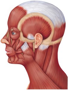
The muscles of the head are responsible for facial expressions, mastication (chewing), and movements of the tongue and eyes. These muscles are unique because many attach to the skin or other muscles rather than bone.
Occipitalis: Moves the scalp back.
Epicranial aponeurosis: A broad tendon connecting the occipitalis and frontalis muscles.
Platysma: Tenses skin of the neck and helps depress the mandible.
Orbicularis oculi: Surrounds the orbit and allows closing of the eyelids.
Orbicularis oris: Encircles the mouth, enabling movements such as puckering.
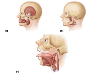
Masseter: Major muscle for jaw elevation during chewing.
Medial and lateral pterygoid: Assist in jaw movements, including side-to-side motion.
Genioglossus, styloglossus, hyoglossus: Muscles that control tongue movement.
Extraocular Muscles
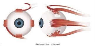
These muscles control the movement of the eyeball and are essential for vision. Each muscle has a specific action, such as moving the eye up, down, or side-to-side.
Superior, inferior, lateral, and medial rectus: Move the eye in the respective directions.
Superior and inferior oblique: Rotate the eye and assist in complex movements.
Common tendinous ring: Origin for several extraocular muscles.
Muscles of the Neck and Vertebral Column
Neck Muscles
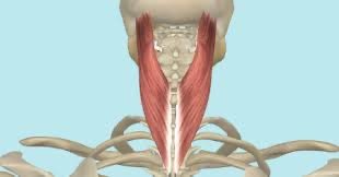
Neck muscles stabilize and move the head and cervical spine. They also assist in swallowing and breathing.
Sternocleidomastoid: Rotates and flexes the neck.
Scalene muscles: Assist in neck flexion and elevate the first two ribs during inspiration.
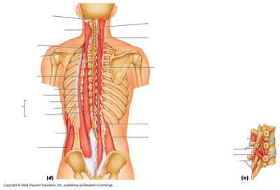
Muscles of the Vertebral Column
These muscles maintain posture and allow movement of the spine. They are organized into superficial, intermediate, and deep layers.
Erector spinae group: Main extensors of the vertebral column.
Multifidus, semispinalis: Stabilize and rotate the vertebral column.
Muscles of the Thorax
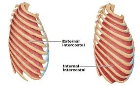
Intercostal Muscles
Intercostal muscles are located between the ribs and are essential for breathing by aiding in the expansion and contraction of the thoracic cavity.
External intercostals: Elevate the ribs during inspiration.
Internal intercostals: Depress the ribs during forced expiration.
Diaphragm
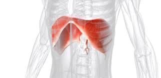
The diaphragm is the primary muscle of respiration, separating the thoracic and abdominal cavities. Its contraction increases thoracic volume, drawing air into the lungs.
Diaphragm: Dome-shaped muscle, contracts to flatten and increase thoracic volume.
Abdominal Wall and Pelvic Floor Muscles
Abdominal Wall Muscles
These muscles support abdominal organs, assist in trunk movement, and help maintain posture.
Transversus abdominis: Compresses abdominal contents.
Internal and external obliques: Rotate and flex the trunk.
Rectus abdominis: Flexes the vertebral column.
Pelvic Floor Muscles
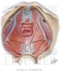
Pelvic floor muscles support pelvic organs and control the openings of the urethra, vagina, and anus.
Pubococcygeus, iliococcygeus: Main muscles of the pelvic floor.
Muscles of the Shoulder, Arm, and Forearm
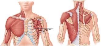
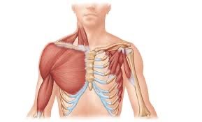
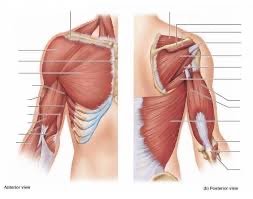
Shoulder Muscles
Muscles crossing the shoulder joint allow for a wide range of arm movements.
Deltoid: Abducts the arm.
Supraspinatus, infraspinatus, teres minor: Rotator cuff muscles stabilize the shoulder.
Latissimus dorsi: Extends, adducts, and medially rotates the arm.
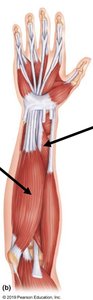
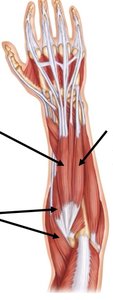
Forearm Muscles
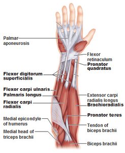
Forearm muscles control movements of the wrist, hand, and fingers. They are divided into flexors and extensors.
Flexor carpi radialis, flexor carpi ulnaris: Flex the wrist.
Extensor carpi radialis longus/brevis: Extend the wrist.
Pronator teres, pronator quadratus: Pronate the forearm.
Muscles of the Thigh and Leg
Thigh Muscles
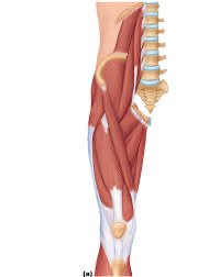
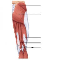
Thigh muscles are responsible for movements of the hip and knee joints. They are grouped into anterior, medial, and posterior compartments.
Quadriceps femoris: Extends the knee.
Gluteus maximus, gluteus medius: Extend and abduct the hip.
Sartorius: Flexes, abducts, and laterally rotates the thigh.
Biceps femoris, semitendinosus, semimembranosus: Hamstring muscles, flex the knee and extend the hip.
Adductors, gracilis, pectineus: Medial compartment, adduct the thigh.
Leg Muscles
Leg muscles control movements of the ankle and toes. They are divided into anterior, posterior, and lateral compartments.
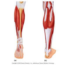
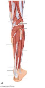
Tibialis anterior: Dorsiflexes the foot.
Gastrocnemius, soleus: Plantar flex the foot.
Peroneus (fibularis) muscles: Evert the foot.
Flexor and extensor hallucis longus: Move the big toe.
Skeletal Muscle Structure and Organization
Connective Tissue Sheaths
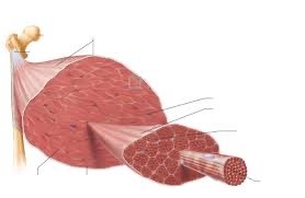
Skeletal muscle is organized into layers of connective tissue that provide support and transmit force.
Endomysium: Surrounds each muscle fiber; composed of fine areolar connective tissue.
Perimysium: Surrounds fascicles (groups of muscle fibers); composed of dense irregular connective tissue.
Epimysium: Surrounds the entire muscle; composed of dense irregular fibrous connective tissue.
Deep fascia: Separates and supports groups of muscles.
Muscle Attachment
Muscles attach to bones via tendons or aponeuroses, spanning joints to produce movement.
Origin: Attachment to the bone that remains stationary during contraction.
Insertion: Attachment to the bone that moves during contraction.
Direct attachment: Epimysium fused to periosteum or perichondrium.
Indirect attachment: Most common, involving tendons or aponeuroses.
Muscle Actions and Interactions
Muscles work together or in opposition to produce movement. They are classified by their roles in movement.
Prime mover (agonist): Main muscle responsible for a movement.
Antagonist: Opposes or reverses a movement.
Synergist: Assists the prime mover, stabilizes joints, or adds force.
Fixator: Immobilizes a bone or muscle origin.
Arrangement of Fascicles
The arrangement of muscle fascicles affects the range of motion and power of a muscle.
Parallel: Fascicles run along the length of the muscle; good range of motion.
Pennate: Short fascicles attach to a central tendon; can be unipennate, bipennate, or multipennate.
Circular: Fascicles arranged in concentric rings; control openings.
Convergent: Fascicles converge toward a single tendon.
Naming Skeletal Muscles
Criteria for Muscle Naming
Skeletal muscles are named based on several criteria, which help identify their location, shape, size, and function.
Location: Named for the bone or body region they are associated with.
Shape: Based on the muscle's shape (e.g., deltoid = triangle).
Size: Terms like maximus (large), minimus (small), longus (long).
Direction of fibers: Rectus (straight), transversus (right angles), oblique (angles).
Number of origins: Bi- (2), tri- (3).
Location of attachments: Named for origin and insertion, with origin listed first.
Action: Named for the action they produce (e.g., flexor, extensor).
Combined criteria: Example: extensor carpi radialis longus.
Summary Table: Muscle Organization and Naming
Criterion | Description | Example |
|---|---|---|
Location | Bone or region | Brachialis (arm) |
Shape | Form of muscle | Deltoid (triangle) |
Size | Relative size | Gluteus maximus |
Direction | Fiber orientation | Rectus abdominis |
Number of origins | Attachment points | Biceps brachii |
Attachment location | Origin/insertion | Sternocleidomastoid |
Action | Movement produced | Flexor carpi radialis |
Additional info: Academic context was added to clarify muscle functions, naming conventions, and anatomical relationships for exam preparation.
