 Back
Study Guide: Spinal Cord, Spinal Nerves, and Reflexes (ANP)
Study Guide - Smart Notes
 Tailored notes based on your materials, expanded with key definitions, examples, and context.
Q1. The spinal cord is vitally important in processing complex information. (True/False)
Background
Topic: Spinal Cord Function
This question tests your understanding of the roles of the spinal cord in the central nervous system, especially regarding information processing and integration.
Key Terms:
Spinal cord: A major part of the central nervous system that transmits signals between the brain and the rest of the body.
Processing complex information: Involves integration, reflexes, and coordination of sensory and motor signals.
Step-by-Step Guidance
Recall the main functions of the spinal cord: conduction, neural integration, locomotion, and reflexes.
Consider whether the spinal cord is responsible for higher-level processing (like the brain) or more basic integration and reflexes.
Think about examples of what the spinal cord can and cannot do in terms of information processing.
Try solving on your own before revealing the answer!
Q2. Each portion of the spinal cord, from which a spinal nerve emerges, is called a "segment" of the cord. (True/False)
Background
Topic: Spinal Cord Anatomy
This question checks your knowledge of spinal cord segmentation and the emergence of spinal nerves.
Key Terms:
Segment: A region of the spinal cord from which a pair of spinal nerves arises.
Spinal nerve: A mixed nerve carrying motor, sensory, and autonomic signals between the spinal cord and the body.
Step-by-Step Guidance
Recall how the spinal cord is divided anatomically (cervical, thoracic, lumbar, sacral, coccygeal).
Think about how spinal nerves are named and where they emerge from the spinal cord.
Determine if each pair of spinal nerves corresponds to a specific segment of the cord.
Try solving on your own before revealing the answer!
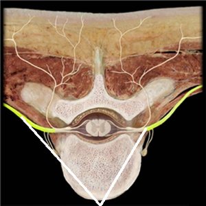
Q47. Which structures are highlighted in the picture below (reference leader lines)?
Background
Topic: Spinal Nerve Anatomy
This question tests your ability to identify anatomical structures of the spinal nerves in a cross-sectional image.
Key Terms:
Dorsal root ganglia: Clusters of sensory neuron cell bodies located in the dorsal root of a spinal nerve.
Ventral rootlets: Small bundles of motor neuron axons that emerge from the anterior side of the spinal cord.
Step-by-Step Guidance
Examine the image for the location of the highlighted structures relative to the spinal cord and vertebra.
Recall the anatomical position of dorsal (posterior) and ventral (anterior) roots and ganglia.
Match the highlighted structures to their correct anatomical names based on their position and appearance.
Try solving on your own before revealing the answer!
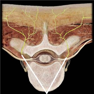
Q48. Which spinal nerve branches are highlighted (reference leader lines)?
Background
Topic: Spinal Nerve Branches
This question asks you to identify specific branches of spinal nerves in a cross-sectional image.
Key Terms:
Ventral rami: Branches of spinal nerves that supply the anterior and lateral trunk and limbs.
Dorsal rami: Branches that supply the muscles and skin of the back.
Step-by-Step Guidance
Look at the image and note the direction and location of the highlighted branches.
Recall the anatomical course of ventral and dorsal rami after the spinal nerve exits the vertebral column.
Use the position of the branches to determine which are ventral and which are dorsal.
Try solving on your own before revealing the answer!
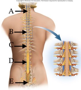
Q73.1 Where do the nerves that innervate the lower limbs go in/out of the spinal cord?
Background
Topic: Spinal Cord Regions and Innervation
This question tests your understanding of the anatomical regions of the spinal cord and which regions give rise to nerves that innervate the lower limbs.
Key Terms:
Lumbosacral enlargement: The region of the spinal cord where nerves to the lower limbs arise.
Cauda equina: A bundle of nerve roots extending from the lower end of the spinal cord.
Step-by-Step Guidance
Examine the labeled diagram and identify the regions marked A–E.
Recall which region of the spinal cord is responsible for lower limb innervation.
Match the correct letter to the lumbosacral enlargement or cauda equina region.
Try solving on your own before revealing the answer!
