Back
BackStudy Guide: The Appendicular Skeleton (Chapter 7)
Study Guide - Smart Notes
 Tailored notes based on your materials, expanded with key definitions, examples, and context.
The Appendicular Skeleton
Overview of the Appendicular Skeleton
The appendicular skeleton consists of the bones that support the limbs and enable movement. It is divided into the pectoral girdle, upper limbs, pelvic girdle, and lower limbs. These structures are essential for locomotion and manipulation of the environment.
Pectoral girdle: Clavicle and scapulae
Upper limbs: Humerus, ulna, radius, carpals, metacarpals, phalanges
Pelvic girdle: Coxal bones (ilium, ischium, pubis)
Lower limbs: Femur, tibia, fibula, patella, tarsals, metatarsals, phalanges
Axial vs. Appendicular Skeleton
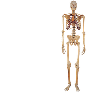
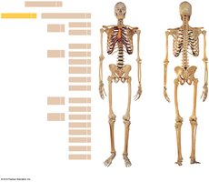
The human skeleton is divided into two main parts: the axial skeleton and the appendicular skeleton. The axial skeleton consists of 80 bones, while the appendicular skeleton consists of 126 bones.
Axial skeleton: Skull, vertebral column, rib cage
Appendicular skeleton: Limbs and girdles
Region | Number of Bones |
|---|---|
Axial Skeleton | 80 |
Appendicular Skeleton | 126 |
Pectoral Girdle | 4 |
Upper Limbs | 60 |
Pelvic Girdle | 2 |
Lower Limbs | 60 |
The Pectoral Girdle and Upper Limb
The Clavicle
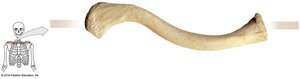
The clavicle, or collarbone, is a slender, S-shaped bone that connects the pectoral girdle to the axial skeleton. It has two ends:
Sternal end: Medial; articulates with the sternum
Acromial end: Lateral; articulates with the scapula
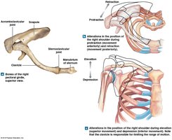
The Scapula
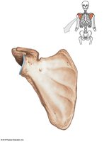
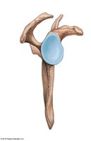
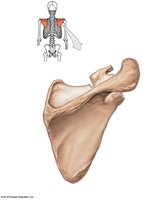
The scapula, or shoulder blade, is a flat, triangular bone that forms the socket of the shoulder joint. The glenoid cavity is the site of articulation with the humerus.
Key features: Acromion, coracoid process, glenoid cavity, spine, borders (superior, medial, lateral), angles (superior, inferior, lateral)
The Upper Limb Bones
The upper limb consists of the humerus, ulna, radius, carpal bones, metacarpals, and phalanges. These bones allow for a wide range of motion and dexterity.
Humerus: The bone of the upper arm
Ulna and Radius: Forearm bones
Carpal bones: Eight bones of the wrist
Metacarpals: Five bones of the palm
Phalanges: Fourteen bones of the fingers
The Humerus
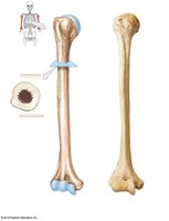
The humerus is the largest bone of the upper limb. It articulates proximally with the scapula and distally with the radius and ulna.
Proximal structures: Head, greater and lesser tubercles, anatomical and surgical necks, intertubercular sulcus
Shaft: Deltoid tuberosity, radial groove
Distal structures: Capitulum, trochlea, epicondyles, fossae (radial, coronoid, olecranon)
The Ulna and Radius
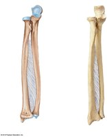
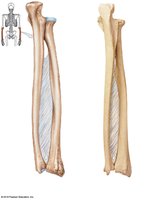
The ulna and radius are the two bones of the forearm. The ulna is medial, and the radius is lateral in anatomical position. They are connected by the interosseous membrane.
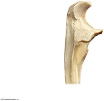
Ulna: Olecranon, trochlear notch, radial notch, ulnar styloid process
Radius: Head, radial tuberosity, radial styloid process
Interosseous membrane: Fibrous sheet between the ulna and radius
The Carpal Bones
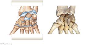
The wrist contains eight carpal bones, arranged in two rows:
Proximal row: Scaphoid, lunate, triquetrum, pisiform
Distal row: Trapezium, trapezoid, capitate, hamate
The Metacarpals and Phalanges
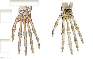
The metacarpals are numbered I to V, starting from the thumb (pollex). Each hand has 14 phalanges: proximal, middle, and distal for each finger except the thumb, which has only proximal and distal phalanges.
Metacarpal I: Associated with the thumb
Phalanges: Proximal, middle, distal (thumb lacks middle phalanx)
The Pelvic Girdle and Lower Limb
The Pelvic Girdle
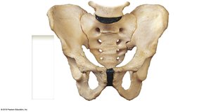
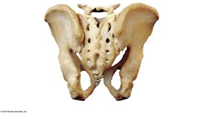
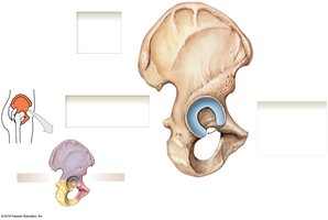
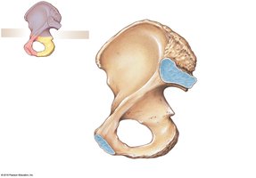
The pelvic girdle consists of two coxal (hip) bones, each formed by the fusion of the ilium, ischium, and pubis. It supports the weight of the upper body and protects pelvic organs.
Ilium: Iliac crest, iliac fossa, anterior/posterior superior/inferior iliac spines
Ischium: Ischial tuberosity, ischial spine, lesser sciatic notch
Pubis: Superior/inferior pubic ramus, pubic symphysis
Acetabulum: Socket for the femur
Obturator foramen: Large opening for nerves and blood vessels
The Pelvis
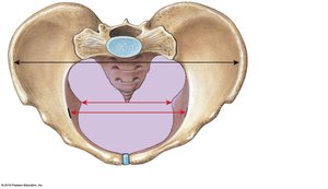
The pelvis is formed by the two coxal bones, sacrum, and coccyx. It is divided into the greater (false) pelvis and lesser (true) pelvis. The pelvic inlet and outlet are important for childbirth.
Greater pelvis: Area between iliac crests
Lesser pelvis: Area below the iliopectineal line
Pelvic inlet: Superior opening
Pelvic outlet: Inferior opening
Male vs. Female Pelvis
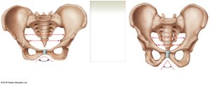
The female pelvis is adapted for childbirth, with a wider pelvic inlet and outlet, less curvature of the sacrum and coccyx, and a broader pubic angle.
Pelvic outlet: Larger in females
Sacrum curve: Less in females
Pelvic inlet: Wider in females
Pubic angle: Greater than 100º in females, less than 90º in males
The Lower Limb Bones
The lower limb consists of the femur, patella, tibia, fibula, tarsal bones, metatarsals, and phalanges. These bones support the body and enable movement.
Femur: Thigh bone, largest bone in the body
Patella: Sesamoid bone protecting the knee joint
Tibia: Medial bone of the leg
Fibula: Lateral bone of the leg
Tarsal bones: Seven bones of the ankle
Metatarsals: Five bones of the foot
Phalanges: Fourteen bones of the toes
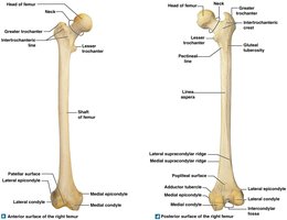
The Femur
The femur is the longest and strongest bone in the body. It articulates proximally with the pelvis and distally with the tibia and patella.
Proximal structures: Head, fovea, neck, greater and lesser trochanters, intertrochanteric line and crest
Distal structures: Medial and lateral condyles, epicondyles, intercondylar fossa, popliteal surface, patellar surface
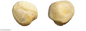
The Patella
The patella is a large sesamoid bone that protects the knee joint. Its anterior surface is rough for tendon attachment, while the posterior surface has concave facets for articulation with the femoral condyles.
Apex: Pointed end
Base: Broad end
Medial and lateral facets: Articulate with femoral condyles
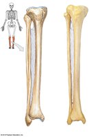
The Tibia and Fibula
The tibia is the main weight-bearing bone of the leg, located medially. The fibula is lateral and provides muscle attachment and stability to the ankle.
Tibia: Tibial tuberosity, medial and lateral condyles, anterior margin, interosseous border, medial malleolus
Fibula: Head, lateral malleolus
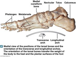
The Tarsal Bones
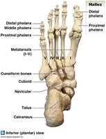
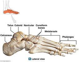
The ankle contains seven tarsal bones:
Calcaneus: Heel bone
Talus: Articulates with tibia and fibula
Navicular, cuboid, medial/intermediate/lateral cuneiform bones: Contribute to the structure of the foot
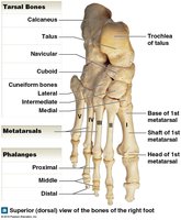
The Metatarsals and Phalanges
The metatarsals are numbered I to V, starting from the hallux (great toe). Each foot has 14 phalanges: proximal, middle, and distal for each toe except the hallux, which has only proximal and distal phalanges.
Metatarsal I: Associated with the hallux
Phalanges: Proximal, middle, distal (hallux lacks middle phalanx)
Arches of the Foot
The foot has two main arches: the longitudinal arch and the transverse arch. These arches help distribute body weight and provide springiness to the step.
Longitudinal arch: Runs from heel to toe
Transverse arch: Runs across the foot