 Back
BackStudy Guide: The Autonomic Nervous System (ANS) – Structure, Function, and Divisions
Study Guide - Smart Notes
 Tailored notes based on your materials, expanded with key definitions, examples, and context.
The Autonomic Nervous System (ANS)
Overview and Organization
The autonomic nervous system (ANS) is a major component of the peripheral nervous system (PNS) responsible for regulating involuntary functions of smooth muscle, cardiac muscle, and glands. It operates largely via subconscious control, ensuring optimal support for body activities such as heart rate, blood pressure, digestion, and pupillary responses.
Motor Neurons: ANS motor neurons innervate visceral organs and tissues.
Control Centers: The hypothalamus, medulla oblongata, pons, cerebral cortex, and limbic system are key regulators.
Divisions: The ANS is divided into the sympathetic and parasympathetic divisions.
Alternate Names: Also known as the involuntary nervous system or visceral motor system.
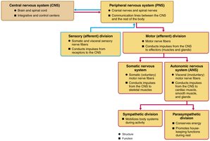
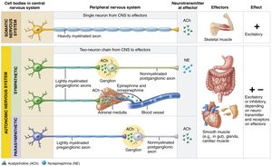
Somatic vs. Autonomic Nervous System
Both the somatic and autonomic nervous systems are regulated by higher brain centers and often work together to adapt to internal and external conditions. However, they differ in their effectors, neural pathways, and neurotransmitter actions.
Effectors:
Somatic Nervous System (SNS): Innervates skeletal muscles.
Autonomic Nervous System (ANS): Innervates cardiac muscle, smooth muscle, and glands.
Number of Neurons:
SNS: Single-neuron pathway from CNS to skeletal muscle.
ANS: Two-neuron chain (preganglionic and postganglionic neurons).
Neurotransmitter Effects:
SNS: Acetylcholine (ACh) is always excitatory.
ANS: Norepinephrine (NE) and ACh can be excitatory or inhibitory, depending on receptor type.

Divisions of the Autonomic Nervous System
Parasympathetic Division
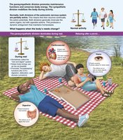
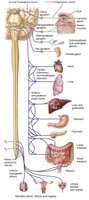
The parasympathetic division is often referred to as the "rest-and-digest" system. It conserves energy and promotes maintenance activities.
Functions:
Reduces heart rate, blood pressure, and respiratory rate.
Increases gastrointestinal activity.
Constricts pupils and accommodates lenses for close vision.
Example: Relaxing after a meal.
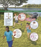
Sympathetic Division
The sympathetic division is known as the "fight-or-flight" system, mobilizing the body during activity, excitement, or emergencies.
Functions:
Increases heart rate, blood pressure, and respiratory rate.
Dilates bronchioles and pupils for far vision.
Shunts blood to skeletal muscles and heart.
Decreases gastrointestinal activity.
Stimulates liver to release glucose.
Example: Vigorous physical activity or emergency situations.
Dual Innervation and Homeostasis
Most visceral organs are served by both divisions, which exert opposite effects to maintain homeostasis through dynamic antagonism.
Anatomical Differences Between ANS Divisions
Key Anatomical Differences
The sympathetic and parasympathetic divisions differ in their sites of origin, fiber lengths, and ganglia locations.
Sites of Origin:
Parasympathetic: Craniosacral (brain and sacral spinal cord).
Sympathetic: Thoracolumbar (thoracic and lumbar spinal cord).
Fiber Lengths:
Parasympathetic: Long preganglionic, short postganglionic fibers.
Sympathetic: Short preganglionic, long postganglionic fibers.
Ganglia Location:
Parasympathetic: In or near effector organs.
Sympathetic: Close to spinal cord.
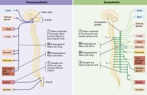
Parasympathetic Division Pathways
Cranial Part: Preganglionic fibers run within cranial nerves III (Oculomotor), VII (Facial), IX (Glossopharyngeal), and X (Vagus). The Vagus nerve accounts for 90% of all preganglionic parasympathetic fibers, innervating organs in the neck, thoracic, and abdominal cavities.
Sacral Part: Preganglionic fibers travel in spinal nerves S2–S4, forming pelvic splanchnic nerves and the inferior hypogastric plexus, supplying the large intestine, urinary bladder, ureters, and reproductive organs.


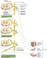
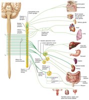
Sympathetic Division Pathways
The sympathetic division is more complex, innervating more organs and some structures exclusively (e.g., sweat glands, arrector pili muscles, blood vessel walls).
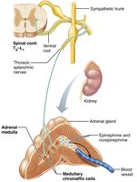
Origin: Cell bodies in spinal cord segments T1–L2 (lateral horn).
Pathways: Preganglionic fibers exit via ventral roots, pass through white ramus communicans, and synapse in sympathetic trunk ganglia.
Trunk Ganglia: Chain ganglia run alongside the vertebral column.
Synapse Options:
Immediate trunk ganglion
Higher or lower trunk ganglion
Collateral ganglion (especially T5 and below)
Splanchnic Nerves: Thoracic, lumbar, and sacral splanchnic nerves form abdominal aortic plexuses (celiac, superior mesenteric, inferior mesenteric ganglia).
Adrenal Medulla Pathway: Some preganglionic fibers pass directly to the adrenal medulla, stimulating chromaffin cells to release epinephrine and norepinephrine into the blood.


ANS Neurotransmitters and Receptors
Major Neurotransmitters
Acetylcholine (ACh): Released by cholinergic fibers (all ANS preganglionic axons, all parasympathetic postganglionic axons).
Norepinephrine (NE): Released by adrenergic fibers (almost all sympathetic postganglionic axons, except sweat glands).
Effect: The action of ACh and NE depends on the receptor type present on the effector organ.
Cholinergic Receptors
Nicotinic Receptors: Found on all postganglionic neurons, adrenal medulla cells, and skeletal muscle cells. ACh binding is always excitatory.
Muscarinic Receptors: Found on all effector cells stimulated by postganglionic cholinergic fibers and some sympathetic targets (e.g., sweat glands). ACh binding can be excitatory or inhibitory, depending on the target organ.
Example: ACh slows heart rate (cardiac muscle) but increases motility (intestinal smooth muscle).
Adrenergic Receptors
Alpha (α) Receptors: α1 and α2 subtypes.
Beta (β) Receptors: β1, β2, and β3 subtypes.
Effect: NE or epinephrine binding can be excitatory or inhibitory, depending on receptor subtype and organ.
NE binding to β1 increases heart rate and blood pressure.
Epinephrine binding to β2 relaxes bronchioles.
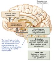
Levels of ANS Control
Hierarchy of Control
The ANS is regulated at multiple levels, with the hypothalamus serving as the main integration center.
Cerebral Cortex: Provides emotional input via the limbic system.
Hypothalamus: Integrates overall ANS activity.
Brain Stem: Regulates pupil size, heart rate, blood pressure, airflow, and salivation.
Spinal Cord: Controls reflexes for urination, defecation, erection, and ejaculation.
Summary Table: Comparison of Somatic and Autonomic Nervous Systems
Feature | Somatic Nervous System | Autonomic Nervous System |
|---|---|---|
Effectors | Skeletal muscle | Cardiac muscle, smooth muscle, glands |
Neural Pathway | Single-neuron from CNS to effector | Two-neuron chain (preganglionic and postganglionic) |
Neurotransmitter | Acetylcholine (ACh) | ACh (parasympathetic), NE (sympathetic) |
Effect | Always excitatory | Excitatory or inhibitory (depends on receptor) |
Key Equations and Concepts
Neurotransmitter Release
Somatic Motor Neuron:
Autonomic Motor Neuron:
Receptor Effects
Nicotinic:
Muscarinic:
Adrenergic:
Conclusion
The autonomic nervous system is essential for maintaining internal balance and responding to environmental changes. Its dual divisions, neurotransmitter systems, and hierarchical control ensure precise regulation of involuntary functions critical for survival and homeostasis.
