 Back
Study Guide: The Axial and Appendicular Skeleton
Study Guide - Smart Notes
 Tailored notes based on your materials, expanded with key definitions, examples, and context.
The Axial Skeleton
Regional Vertebral Characteristics
The vertebral column is divided into cervical, thoracic, lumbar, sacral, and coccygeal regions, each with distinct structural features and functions.
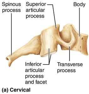
Cervical Vertebrae: Seven vertebrae (C1–C7) located in the neck. The first two, the atlas (C1) and axis (C2), are specialized for head movement.
Atlas (C1): Lacks a body and spinous process; supports the skull and allows nodding motion.
Axis (C2): Has a prominent dens (odontoid process) that acts as a pivot for rotation, enabling side-to-side movement.
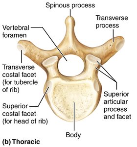
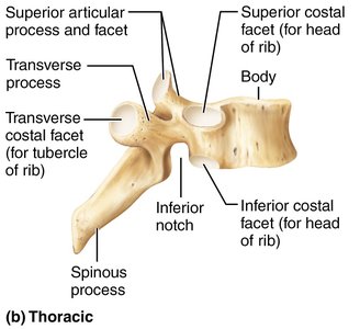
Thoracic Vertebrae: Twelve vertebrae (T1–T12) articulate with ribs; have heart-shaped bodies and circular vertebral foramina.
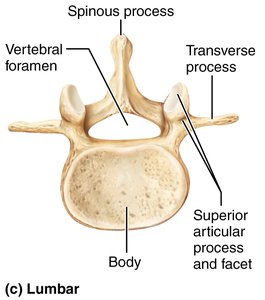
Lumbar Vertebrae: Five vertebrae (L1–L5) in the lower back; have massive, kidney-shaped bodies to bear weight.
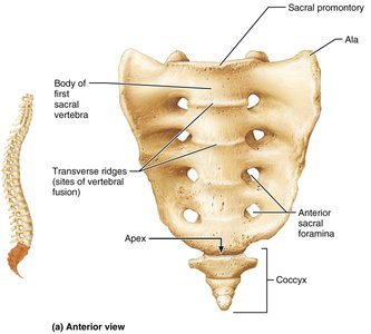
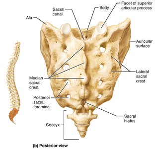
Sacrum: Five fused vertebrae forming the posterior wall of the pelvis; articulates with the hip bones.
Coccyx: Three to five fused vertebrae forming the tailbone; minimal function.
Atlas and Axis
The atlas and axis are the first two cervical vertebrae, crucial for head movement.
Atlas (C1): Supports the skull, allows nodding.
Axis (C2): Contains the dens, which acts as a pivot for rotation.
Movement: The dens enables the atlas to rotate, allowing the head to turn side-to-side.
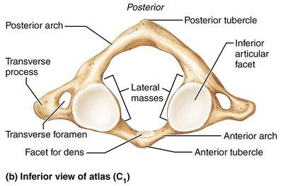
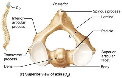
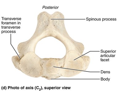
Comparison of Vertebral Regions
Each vertebral region has unique characteristics, summarized in the table below:
Characteristic | Cervical (3–7) | Thoracic | Lumbar |
|---|---|---|---|
Body | Small, oval, wide side to side | Heart-shaped; bears costal facets | Massive; kidney-shaped |
Spinous process | Short; bifid (except C7) | Long; sharp; projects inferiorly | Short; blunt; rectangular |
Vertebral foramen | Triangular, large | Circular | Triangular |
Transverse processes | Contain foramina | Bear facets for ribs (except T11, T12) | Thin and tapered |
Movements allowed | Flexion, extension, lateral flexion, rotation | Rotation, limited flexion/extension | Flexion, extension, some lateral flexion; rotation prevented |
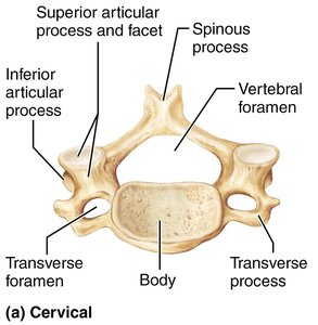

Sacrum and Coccyx
The sacrum and coccyx form the base of the vertebral column and are important for pelvic structure.
Sacrum: Triangular bone, five fused vertebrae, forms sacroiliac joints with hip bones.
Key features: Sacral promontory, transverse ridges, anterior and posterior sacral foramina, alae, median and lateral sacral crests, sacral canal, sacral hiatus.
Coccyx: Three to five fused vertebrae; minimal function.
The Thoracic Cage
The thoracic cage protects vital organs and supports the upper body.
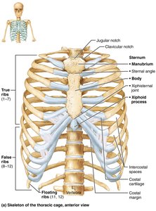
Components: Thoracic vertebrae, ribs, sternum, costal cartilages.
Functions: Protection, support, muscle attachment.
Sternum
The sternum, or breastbone, consists of three fused bones: manubrium, body, and xiphoid process.
Manubrium: Articulates with clavicle and ribs 1–2.
Body: Articulates with ribs 2–7.
Xiphoid process: Site of muscle attachment; not ossified until ~age 40.
Landmarks: Jugular notch, sternal angle, xiphisternal joint.
Ribs
There are 12 pairs of ribs, classified as true, false, and floating ribs.
True ribs (1–7): Attach directly to sternum via costal cartilages.
False ribs (8–10): Attach indirectly to sternum.
Floating ribs (11–12): No attachment to sternum.
Main parts of a rib: Shaft, head, neck, tubercle.
The Appendicular Skeleton
Pectoral Girdle
The pectoral girdle attaches the upper limbs to the axial skeleton and provides mobility.
Components: Clavicles (collarbones) and scapulae (shoulder blades).
Clavicle: S-shaped, anchors muscles, braces scapulae and arms laterally.
Scapula: Thin, triangular bone with three borders (superior, medial, lateral) and three angles (superior, lateral, inferior).
Features: Spine, acromion, coracoid process, suprascapular notch, fossae.
Upper Limb
The upper limb consists of the arm, forearm, and hand.
Arm: Humerus is the only bone; articulates with scapula, radius, and ulna.
Forearm: Radius (lateral) and ulna (medial); connected by interosseous membrane.
Hand: Carpus (wrist, 8 bones), metacarpus (palm, 5 bones), phalanges (fingers, 14 bones).
Pelvic Girdle
The pelvic girdle attaches the lower limbs to the axial skeleton and supports pelvic organs.
Components: Two hip bones (coxal bones) and sacrum.
Coxal bone: Formed by fusion of ilium, ischium, and pubis.
Acetabulum: Deep socket for femur head.
Pelvic Structure and Childbearing
Female pelvis: Wider, shallower, lighter, rounder; adapted for childbearing.
Male pelvis: Narrower, deeper, heavier, more prominent markings.
Pelvic brim: Defines birth canal.
Lower Limb
The lower limb carries the body's weight and is subjected to high forces.
Thigh: Femur is the largest and strongest bone; articulates with hip and knee.
Leg: Tibia (medial, weight-bearing) and fibula (lateral, not weight-bearing).
Foot: Tarsus (7 bones), metatarsus (5 bones), phalanges (14 bones).
Arches: Lateral longitudinal, medial longitudinal, transverse; maintained by bones, ligaments, tendons.
Developmental Aspects of the Skeleton
Infant Skull and Growth
Infant skull: More bones than adult; connected by fontanelles for birth and brain growth.
Growth: Cranium is large at birth; arms and legs grow faster than head and trunk.
Spine: Primary curvatures (thorax, sacrum) are convex at birth; secondary curvatures (cervical, lumbar) develop later.
Aging and Skeletal Changes
Intervertebral discs: Become thinner, less hydrated, less elastic with age.
Height loss: Common by age 55.
Costal cartilages: Ossify, causing rigid thorax.
Bone mass: Decreases, increasing fracture risk.
Clinical Homeostatic Imbalances
Xiphoid process trauma: Can cause hemorrhaging; proper CPR hand placement is crucial.
Colles' fracture: Distal radius fracture from falls.
Carpal tunnel syndrome: Compression of median nerve in wrist.
Bimalleolar fracture: Break in distal tibia and fibula; common sports injury.
Flat foot (pes planus): May be congenital or acquired; proper footwear helps.
Cleft palate: Failure of palate fusion; interferes with feeding.
Developmental dysplasia of the hip (DDH): Dislocation or poorly developed acetabulum; requires early treatment.
Summary Table: Comparison of Male and Female Pelves
Characteristic | Female | Male |
|---|---|---|
General structure | Tilted forward; adapted for childbearing | Tilted less far forward; adapted for support |
Bone thickness | Lighter, thinner, smoother | Heavier, thicker, more prominent markings |
Acetabula | Smaller, farther apart | Larger, closer together |
Pubic arch | Broader (80–90°) | More acute (50–60°) |
Sacrum | Wider, shorter, less curvature | Narrow, longer, more curvature |
Coccyx | More movable, projects inferiorly | Less movable, projects anteriorly |
Pelvic inlet | Wider, oval | Narrow, heart-shaped |
Pelvic outlet | Wider, ischial spines farther apart | Narrower, ischial spines closer together |
Key Terms
Vertebral foramen: Opening for spinal cord.
Spinous process: Posterior projection for muscle attachment.
Transverse process: Lateral projection for muscle and ligament attachment.
Facet: Smooth surface for articulation.
Acetabulum: Hip socket for femur head.
Fontanelle: Soft spot in infant skull.
Important Formulas
Bone Count:
Examples and Applications
Clinical application: Understanding vertebral structure is essential for diagnosing spinal injuries.
Childbirth: Pelvic structure determines ease of delivery.
Sports medicine: Knowledge of limb bones aids in treating fractures and injuries.
