 Back
BackStudy Guide: The Axial Skeleton (Chapter 7, Anatomy & Physiology)
Study Guide - Smart Notes
 Tailored notes based on your materials, expanded with key definitions, examples, and context.
The Axial Skeleton
Overview of the Axial Skeleton
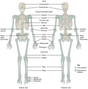
The axial skeleton forms the central axis of the human body and consists of 80 bones, accounting for approximately 40% of the total bones. It includes the skull, vertebral column, and thoracic cage, providing structural support and protection for vital organs.
Skull: 22 bones (8 cranial, 14 facial)
Bones associated with the skull: 6 auditory ossicles, 1 hyoid bone
Thoracic cage: Sternum and 24 ribs
Vertebral column: 24 vertebrae, sacrum, coccyx
The Skull
Structure and Function
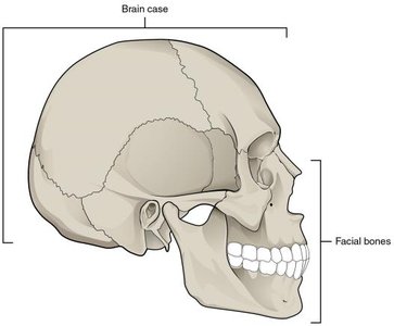
The skull protects the brain and forms the structure of the face. It is divided into the brain case (cranial bones) and facial bones, which together create the cranial cavity, orbits, nasal cavities, and jaws.
Brain case: Encases and protects the brain
Facial bones: Form the upper/lower jaws, nasal cavities, and orbits
Cranial Bones
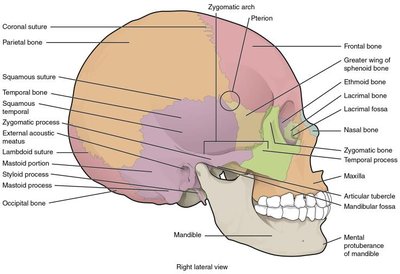
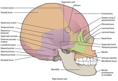
The cranial bones form the brain case and are joined by immobile joints called sutures. Key cranial bones include:
Parietal bones: Form most of the upper lateral side of the skull; joined at the top, bounded by frontal, temporal, and occipital bones
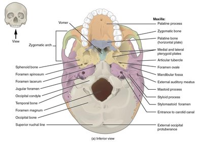
Temporal bones: Form the lower lateral side; include the mastoid process (muscle attachment), external acoustic meatus (ear canal), and mandibular fossa (part of the temporomandibular joint)
Frontal bone: Forms the forehead and floor of the anterior cranial cavity
Occipital bone: Forms the posterior skull and base; contains the foramen magnum (spinal cord exit) and occipital condyles (articulate with C1)
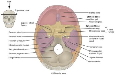
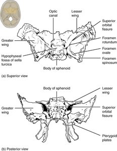
Sphenoid bone: Forms part of the cranial floor, acts as a cross-bridge, unites cranial and facial bones, contains optic canals and sella turcica (pituitary gland housing)
Ethmoid bone: Forms part of the anterior cranial floor, roof of the nasal cavity, part of the nasal septum, and medial orbit wall; includes cribriform plate (olfactory foramina), perpendicular plate, crista galli, and ethmoidal labyrinth
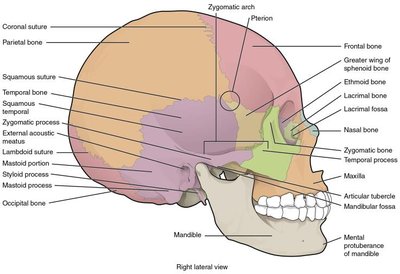
Sutures of the Skull
Sutures are immobile joints between adjacent skull bones. Major sutures include:
Coronal suture: Frontal to parietal bones
Sagittal suture: Right and left parietal bones
Lambdoid suture: Occipital to parietal and temporal bones
Squamous suture: Temporal to parietal bones
Facial Bones
The facial bones form the structure of the face, jaws, and nasal cavities. There are 14 facial bones, including:
Maxillary bones: Form the upper jaw, most of the hard palate, medial floor of the orbit, and lateral base of the nose
Palatine bones: Form the posterior hard palate
Zygomatic bones: Cheekbones; form lateral wall and margin of the orbit
Nasal bones: Form the bridge of the nose
Lacrimal bones: Form the anterior, medial wall of the orbit
Inferior nasal conchae: Project into the nasal cavity; create air turbulence
Vomer: Forms the posterior-inferior part of the nasal septum
Mandible: Forms the lower jaw; only moveable skull bone
Orbit and Nasal Structures
The orbit is a bony socket housing the eyeball, formed by seven skull bones. The nasal septum is composed of bone and cartilage, and the nasal conchae create air turbulence for warming and humidifying inhaled air.
Orbit: Frontal, zygomatic, maxilla, palatine, ethmoid, lacrimal, sphenoid bones
Nasal septum: Perpendicular plate of ethmoid, vomer, septal cartilage
Nasal conchae: Superior, middle (ethmoid), and inferior conchae
Cranial Fossae and Foramina
The floor of the cranial cavity is divided into three fossae: anterior, middle, and posterior. Each contains openings (foramina) for nerves and blood vessels.
Anterior cranial fossa: Contains frontal lobes
Middle cranial fossa: Contains temporal lobes; includes optic canal, foramen rotundum, foramen ovale
Posterior cranial fossa: Contains cerebellum; includes internal acoustic meatus, hypoglossal canal, jugular foramen
Paranasal Sinuses and Hyoid Bone
Paranasal sinuses are air-filled spaces within certain skull bones, lined with mucous epithelium. The hyoid bone is a U-shaped bone in the neck, not connected to other bones, serving as a muscle attachment site.
Sinuses: Frontal, sphenoid, ethmoid, maxillary bones
Functions: Warm/humidify air, trap dust/microbes
Hyoid bone: Attachment for muscles of larynx, pharynx, tongue
The Vertebral Column
Structure and Regions
The vertebral column, or spine, consists of 26 bones: 24 vertebrae, the sacrum, and the coccyx. It provides support, protects the spinal cord, and allows flexibility.
Cervical vertebrae (C1–C7): Neck region; C1 (atlas) and C2 (axis) allow head movement
Thoracic vertebrae (T1–T12): Superior back; articulate with ribs
Lumbar vertebrae (L1–L5): Inferior back; largest, bear most weight
Sacrum: Fusion of 5 vertebrae; part of pelvis
Coccyx: Fusion of 3–5 vertebrae; tailbone
General Vertebral Structure
Each vertebra has a vertebral body (anterior, weight-bearing), vertebral arch (posterior), vertebral foramen (spinal cord passage), and processes for muscle attachment.
Intervertebral foramen: Passageway for spinal nerves
Intervertebral disc: Fibrocartilage, shock absorbing
Transverse and spinous processes: Muscle attachment
Articular processes: Superior and inferior, for articulation between vertebrae
Specialized Vertebrae
Cervical vertebrae have small bodies and transverse foramina for blood vessels. The atlas (C1) lacks a body/spinous process and allows nodding. The axis (C2) has the dens for head rotation. Thoracic vertebrae have costal facets for rib articulation. Lumbar vertebrae are largest and thickest.
Sacrum and Coccyx
The sacrum and coccyx are formed by fusion of vertebrae. The sacrum has transverse ridges, sacral canal, sacral hiatus, and sacral foramina for nerves. The coccyx articulates with the sacrum and may fuse completely in old age.
The Thoracic Cage and Ribs
Structure and Function
The thoracic cage protects the heart and lungs and supports breathing. It consists of the sternum, 12 pairs of ribs, costal cartilages, and thoracic vertebrae.
Sternum: Manubrium (superior, articulates with clavicles and 1st ribs), body (articulates with ribs 2–7), xiphoid process (inferior, muscle attachment)
Ribs: Elongate, curved bones; posteriorly articulate with thoracic vertebrae
True ribs (vertebrosternal): Rib pairs 1–7, direct connection to sternum via costal cartilage
False ribs (vertebrochondral): Rib pairs 8–10, indirect connection via cartilage of rib 7
Floating ribs (vertebral): Rib pairs 11–12, no connection to sternum
Summary Table: Axial Skeleton Components
Region | Main Bones | Key Functions |
|---|---|---|
Skull | Cranial (8), Facial (14), Auditory ossicles (6), Hyoid (1) | Protects brain, forms face, supports sensory organs |
Vertebral Column | Cervical (7), Thoracic (12), Lumbar (5), Sacrum (1), Coccyx (1) | Supports body, protects spinal cord, allows movement |
Thoracic Cage | Sternum (1), Ribs (24) | Protects heart/lungs, supports breathing |
