 Back
BackStudy Guide: The Brain and Cranial Nerves (Anatomy & Physiology) CH14
Study Guide - Smart Notes
 Tailored notes based on your materials, expanded with key definitions, examples, and context.
The Brain and Cranial Nerves
Overview of the Brain
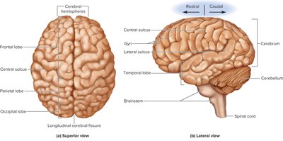
The brain is the central organ of the nervous system, responsible for integrating sensory information, coordinating motor functions, and facilitating higher cognitive processes. It is divided into several major regions, each with distinct anatomical landmarks and functions.
Directional Terms: Rostral (toward the forehead), Caudal (toward the spinal cord).
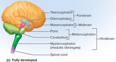
Major Portions: Forebrain, cerebellum, and brainstem.
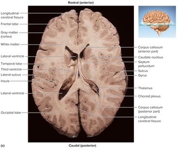
Cerebrum: Largest part of the forebrain, comprising 83% of brain volume. Features include cerebral hemispheres, gyri (thick folds), sulci (shallow grooves), and the longitudinal cerebral fissure.
Corpus Callosum: Thick bundle of nerve fibers connecting the two hemispheres.
Cerebellum: Second-largest part, located in the posterior cranial fossa, separated from the cerebrum by the transverse cerebral fissure.
Brainstem: Includes the midbrain, pons, and medulla oblongata.
Gray and White Matter
The brain is composed of two types of tissue: gray matter and white matter, each with distinct roles in neural processing and communication.
Gray Matter: Contains neuron cell bodies, dendrites, and synapses. Forms the cortex (surface layer) and nuclei (deeper masses).
White Matter: Composed of myelinated axons organized into tracts, connecting different regions of the brain and spinal cord.
Embryonic Development of the CNS
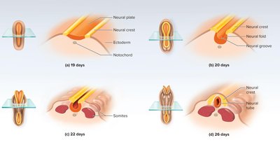
The central nervous system develops from the ectodermal layer of the embryo through a process called neurulation. This process forms the neural tube, which later differentiates into the brain and spinal cord.
Neurulation: Formation of the neural plate, neural groove, and neural tube (by day 26).
Neural Crest: Gives rise to meninges, peripheral nervous system, and other structures.
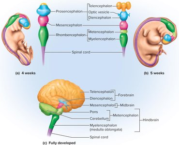
Primary Vesicles (Week 4): Forebrain (prosencephalon), midbrain (mesencephalon), hindbrain (rhombencephalon).
Secondary Vesicles (Week 5): Telencephalon, diencephalon, mesencephalon, metencephalon, myelencephalon.
Meninges, Ventricles, Cerebrospinal Fluid, and Blood Supply
Meninges
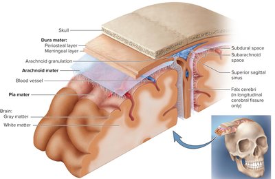
The meninges are three protective membranes surrounding the brain and spinal cord, providing structural support and protection.
Dura Mater: Outermost, tough layer; consists of periosteal and meningeal layers.
Arachnoid Mater: Transparent membrane; subarachnoid space contains cerebrospinal fluid (CSF).
Pia Mater: Innermost, delicate layer; closely follows brain contours.
Dural Sinuses: Spaces between dura layers that collect venous blood.
Dural Folds: Falx cerebri, tentorium cerebelli, falx cerebelli separate brain regions.
Ventricles and Cerebrospinal Fluid (CSF)
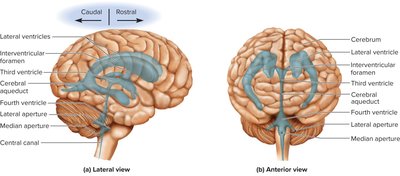
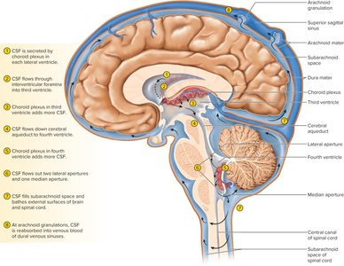
The brain contains four internal ventricles filled with CSF, which cushions and nourishes neural tissue.
Ventricles: Two lateral ventricles, third ventricle, fourth ventricle; connected by interventricular foramen and cerebral aqueduct.
CSF Production: Produced by choroid plexus and ependymal cells; flows through ventricles and subarachnoid space.
Functions: Buoyancy, protection, chemical stability.
Blood Supply and Brain Barrier System
The brain receives a significant portion of the body's blood supply and is protected by specialized barriers that regulate the entry of substances.
Blood-Brain Barrier: Tight junctions between endothelial cells prevent harmful substances from entering brain tissue.
Blood-CSF Barrier: Tight junctions between ependymal cells at the choroid plexus.
Circumventricular Organs: Areas where the barrier is absent, allowing the brain to monitor blood variables.
Stroke: Sudden death of brain tissue due to blood supply interruption; can be hemorrhagic or ischemic.
The Hindbrain and Midbrain
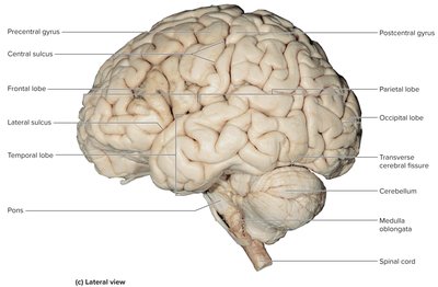
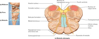
Medulla Oblongata
The medulla oblongata is the most caudal part of the brainstem, responsible for vital autonomic functions and relaying sensory and motor signals.
Anatomical Features: Pyramids, olives, cranial nerve origins, gracile and cuneate fasciculi.
Functions: Cardiac, vasomotor, and respiratory centers; relay for sensory and motor pathways.
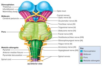
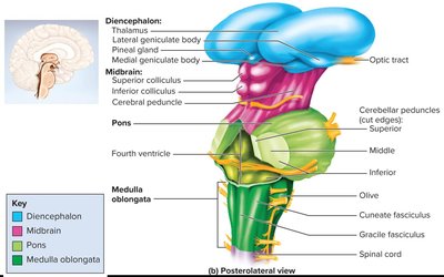
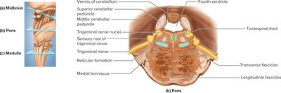
Pons
The pons is a prominent bulge in the brainstem, connecting the medulla to the midbrain and cerebellum. It contains nuclei involved in sensory and motor functions.
Cerebellar Peduncles: Connect pons to cerebellum.
Cranial Nerves: V, VI, VII, VIII.
Functions: Sleep, respiration, posture, facial sensation, and movement.
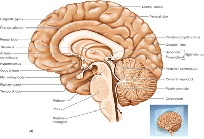
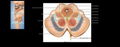
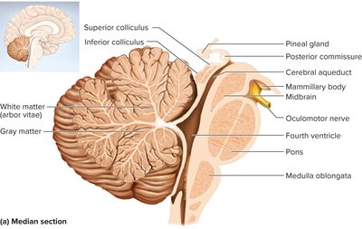
Midbrain
The midbrain connects the hindbrain to the forebrain and contains important nuclei for motor control, pain awareness, and sensory processing.
Tectum: Contains superior and inferior colliculi for visual and auditory reflexes.
Cerebral Peduncles: Include tegmentum (red nucleus), substantia nigra (motor control), and cerebral crus (corticospinal tracts).
Cranial Nerves: III (oculomotor), IV (trochlear).

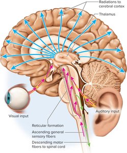
Reticular Formation
The reticular formation is a network of gray matter extending through the brainstem, involved in motor control, pain modulation, sleep, consciousness, and habituation.
Somatic Motor Control: Maintains muscle tone, balance, and posture.
Pain Modulation: Blocks pain signals in the spinal cord.
Sleep and Consciousness: Central role in alertness and sleep; injury can cause coma.
Habituation: Filters repetitive stimuli.
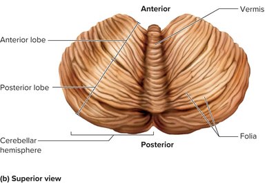
Cerebellum
The cerebellum is the largest part of the hindbrain, crucial for motor coordination and various cognitive functions.
Structure: Two hemispheres connected by vermis; cortex of gray matter, arbor vitae (white matter), deep nuclei.
Cerebellar Peduncles: Inferior (medulla), middle (pons), superior (midbrain).
Functions: Motor coordination, sensory processing, timekeeping, emotion, planning.
The Forebrain
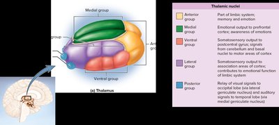
Diencephalon
The diencephalon is a central region of the forebrain, enclosing the third ventricle and comprising the thalamus, hypothalamus, and epithalamus.
Thalamus: Gateway to the cerebral cortex; processes and relays sensory and motor signals; involved in memory and emotion.
Hypothalamus: Major control center for autonomic and endocrine systems; regulates homeostasis, hormone secretion, thermoregulation, food/water intake, sleep, memory, and emotional behavior.
Epithalamus: Contains pineal gland (endocrine) and habenula (relay from limbic system).
Cerebrum
The cerebrum is the largest part of the brain, responsible for higher cognitive functions, sensory perception, voluntary motor actions, and memory.
Lobes: Frontal (motor, planning, emotion), parietal (sensory integration), occipital (vision), temporal (hearing, memory, emotion), insula (language, taste, visceral integration).
White Matter: Projection, commissural, and association tracts connect different brain regions.
Cerebral Cortex: Gray matter layer with stellate and pyramidal cells; neocortex is the most evolved part.
Limbic System: Emotion and learning; includes cingulate gyrus, hippocampus, amygdala.
Basal Nuclei: Motor control; includes caudate nucleus, putamen, globus pallidus.
Integrative Functions of the Brain
Electroencephalogram (EEG) and Brain Waves
EEG records brain waves, which reflect the electrical activity of the cortex. Different types of waves correspond to various mental states.
Alpha Waves: Awake, resting, eyes closed.
Beta Waves: Mental activity, sensory stimulation.
Theta Waves: Drowsy or sleeping adults.
Delta Waves: Deep sleep.
Sleep
Sleep is a reversible loss of consciousness, occurring in distinct stages and regulated by interactions among brain regions.
Stages: 1 (drowsy), 2 (light sleep), 3 (moderate/deep sleep), 4 (deep sleep), REM (rapid eye movement).
REM Sleep: Vivid dreams, EEG resembles waking state, sleep paralysis.
Regulation: Suprachiasmatic nucleus (SCN), pineal gland (melatonin), orexins (wakefulness).
Cognition, Memory, and Emotion
Cognition encompasses mental processes such as perception, reasoning, and judgment. Memory involves learning, storage, and retrieval, with the hippocampus, cerebellum, and amygdala playing key roles. Emotion is regulated by the prefrontal cortex, hypothalamus, and amygdala.
Memory Types: Explicit (facts, events), implicit (skills).
Amnesia: Anterograde (cannot store new info), retrograde (cannot recall old info).
Emotion: Amygdala (fear, attention), hypothalamus (autonomic responses).
Sensation and Motor Control
Sensory input is processed in primary sensory cortices and association areas. Motor control involves the motor association area, precentral gyrus, basal nuclei, and cerebellum.
Special Senses: Vision (occipital lobe), hearing (temporal lobe), equilibrium (cerebellum), taste (postcentral gyrus), smell (temporal lobe).
General Senses: Touch, pressure, pain, temperature.
Motor Homunculus: Diagram of motor cortex representation of body regions.
Basal Nuclei: Initiation and cessation of movement; disorders cause dyskinesias.
Cerebellum: Motor coordination, learning, posture.
Language and Cerebral Lateralization
Language is processed in specialized areas: Wernicke (comprehension), Broca (speech production), and affective language area (emotion). Cerebral lateralization refers to functional differences between hemispheres.
Left Hemisphere: Language, analytical reasoning.
Right Hemisphere: Imagination, spatial relationships, artistic skills.
Lateralization: Correlated with handedness, age, and sex.
The Cranial Nerves
Overview and Classification
The brain communicates with the body via 12 pairs of cranial nerves, each with specific sensory, motor, or mixed functions.
Origin: Most arise from the brainstem; exit through foramina.
Classification: Sensory (I, II, VIII), motor (III, IV, VI, XI, XII), mixed (V, VII, IX, X).
Pathways: Most fibers are ipsilateral; exceptions include optic and trochlear nerves.
Selected Cranial Nerves
Olfactory (I): Sensory for smell.
Optic (II): Sensory for vision.
Oculomotor (III), Trochlear (IV), Abducens (VI): Motor for eye movement.
Trigeminal (V): Largest, sensory and motor for face.
Facial (VII): Motor for facial muscles, sensory for taste.
Vestibulocochlear (VIII): Sensory for hearing and equilibrium.
Glossopharyngeal (IX): Sensory for tongue/throat, motor for swallowing.
Vagus (X): Extensive distribution; sensory and motor for viscera.
Accessory (XI): Motor for head/neck/shoulder.
Hypoglossal (XII): Motor for tongue.
Cranial Nerve Disorders
Trigeminal Neuralgia: Intense facial pain.
Bell Palsy: Facial muscle weakness.
Imaging and Functional Studies
Modern imaging techniques such as PET and fMRI allow visualization of brain activity and blood flow during cognitive tasks.
PET: Uses radioactively labeled glucose to highlight active areas.
fMRI: Measures blood flow and oxygenation changes.
Summary Table: Major Brain Regions and Functions
Region | Main Functions |
|---|---|
Cerebrum | Sensory perception, cognition, voluntary motor actions |
Cerebellum | Motor coordination, balance, learning |
Brainstem | Autonomic functions, relay of signals |
Diencephalon | Homeostasis, hormone regulation, sensory relay |
Limbic System | Emotion, memory |
Basal Nuclei | Motor control |
