 Back
Study Guide: The Brain and Cranial Nerves (Anatomy & Physiology)
Study Guide - Smart Notes
 Tailored notes based on your materials, expanded with key definitions, examples, and context.
The Brain and Cranial Nerves
Overview of Brain Organization
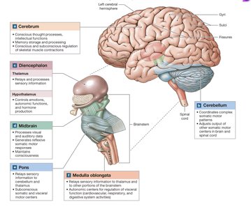
The brain is the central organ of the nervous system, responsible for integrating sensory and motor information, and higher mental functions such as intelligence, memory, and emotions. It is divided into four major regions: the cerebrum, cerebellum, diencephalon, and brainstem.
Cerebrum: Controls conscious thought, intellect, memory, and complex movements.
Cerebellum: Coordinates complex motor patterns and maintains balance.
Diencephalon: Integrates sensory information and controls autonomic functions.
Brainstem: Relays information between the brain and spinal cord; regulates vital functions.
Embryology and Development of the Brain
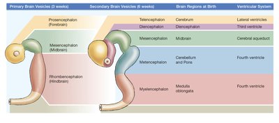
The brain develops from the neural tube, forming three primary brain vesicles: prosencephalon (forebrain), mesencephalon (midbrain), and rhombencephalon (hindbrain). These further differentiate into secondary vesicles and mature brain regions.
Prosencephalon: Forms the telencephalon (cerebrum) and diencephalon (thalamus, hypothalamus).
Mesencephalon: Becomes the midbrain.
Rhombencephalon: Forms the metencephalon (cerebellum and pons) and myelencephalon (medulla oblongata).
Ventricular System of the Brain
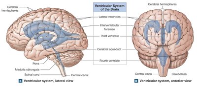
The brain contains four ventricles: two lateral ventricles, the third ventricle, and the fourth ventricle. These are interconnected by passageways and filled with cerebrospinal fluid (CSF), which cushions and supports the brain.
Lateral ventricles: Located in each cerebral hemisphere.
Third ventricle: Located in the diencephalon.
Fourth ventricle: Located between the pons and cerebellum, extending into the medulla oblongata.
Protection and Support of the Brain
Cranial Meninges
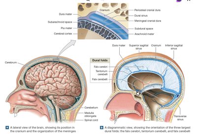
The brain is protected by three layers of cranial meninges: dura mater, arachnoid mater, and pia mater. These layers provide structural support and help maintain the environment of the CNS.
Dura mater: Tough, outermost layer; forms dural folds for stabilization.
Arachnoid mater: Middle layer; contains the subarachnoid space filled with CSF.
Pia mater: Innermost layer; closely adheres to the brain surface.
Cerebrospinal Fluid (CSF)
CSF is produced by the choroid plexus in each ventricle and circulates through the ventricular system and subarachnoid space. It supports the brain, cushions neural structures, and transports nutrients and wastes.
Functions: Support, cushioning, and chemical transport.
Production: Choroid plexus (specialized ependymal cells).
Clinical relevance: Disruption can cause hydrocephalus or other CNS disorders.
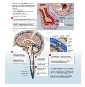
Blood-Brain Barrier
The blood-brain barrier (BBB) isolates CNS tissue from general circulation, restricting passage of substances. It is formed by tight junctions between capillary endothelial cells and supported by astrocytes.
Allows: Lipid-soluble substances (O2, CO2, steroids).
Restricts: Water-soluble substances, ions, and large molecules.
Clinical relevance: Some drugs cannot cross the BBB; exceptions exist in circumventricular organs.
Brainstem Structure and Function
Medulla Oblongata
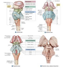
The medulla oblongata connects the brain to the spinal cord and contains nuclei for autonomic functions, cranial nerves, and relay stations.
Autonomic centers: Cardiovascular and respiratory regulation.
Sensory/motor nuclei: Cranial nerves VIII-XII.
Relay stations: Gracile and cuneate nuclei, decussation of pyramids.
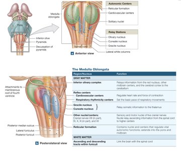
Region/Nucleus | Function |
|---|---|
Reticular formation | Autonomic and reflex activity |
Cardiovascular centers | Heart rate and blood flow regulation |
Respiratory rhythmicity centers | Basic respiratory rhythm |
Gracile/cuneate nuclei | Relay somatic sensory info to thalamus |
Inferior olivary complex | Relay somatic motor commands to cerebellum |
Pons
The pons links the cerebellum with the brainstem, cerebrum, and spinal cord. It contains nuclei for cranial nerves, respiratory control, and relay tracts.
Cranial nerve nuclei: V, VI, VII, VIII.
Respiratory centers: Apneustic and pneumotaxic centers.
Relay tracts: Ascending, descending, and transverse pontine fibers.
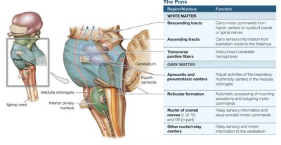
Region/Nucleus | Function |
|---|---|
Descending tracts | Carry motor commands |
Ascending tracts | Sensory info to thalamus |
Transverse pontine fibers | Interconnect cerebellar hemispheres |
Reticular formation | Automatic processing |
Cranial nerve nuclei | Sensory/motor info to/from face |
Midbrain
The midbrain regulates visual and auditory reflexes and controls alertness. It contains sensory nuclei, motor nuclei, and fiber tracts.
Corpora quadrigemina: Superior colliculi (visual), inferior colliculi (auditory).
Red nucleus: Subconscious motor commands.
Substantia nigra: Dopamine production, inhibits basal nuclei.
Cerebral peduncles: Motor and sensory fiber tracts.
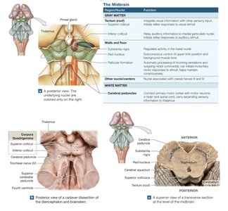
Region/Nucleus | Function |
|---|---|
Superior colliculus | Visual reflexes |
Inferior colliculus | Auditory reflexes |
Red nucleus | Motor coordination |
Substantia nigra | Dopamine, inhibits basal nuclei |
Cerebellum Structure and Function
Cerebellar Anatomy
The cerebellum coordinates reflexive and learned patterns of muscular activity. It consists of two hemispheres, the vermis, and the flocculonodular lobe. The cerebellar cortex contains Purkinje cells, and the arbor vitae connects cortex and nuclei.
Folia: Surface folds.
Purkinje cells: Highly branched neurons for integration.
Arbor vitae: Branching white matter.
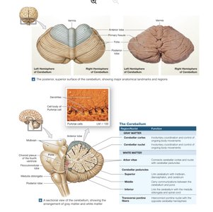
Region/Nucleus | Function |
|---|---|
Cerebellar cortex | Coordination of movement |
Purkinje cells | Integration of sensory/motor info |
Arbor vitae | Connects cortex and nuclei |
Cerebellar Peduncles
Superior: Link cerebellum with midbrain, diencephalon, cerebrum.
Middle: Connect cerebellum with pons.
Inferior: Communicate with medulla oblongata and spinal cord.
Diencephalon Structure and Function
Thalamus
The thalamus is the final relay point for sensory information ascending to the cerebral cortex. It acts as a filter and coordinates activities between the basal nuclei and cortex.
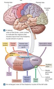
Nuclei/Body | Function |
|---|---|
Anterior nuclei | Limbic system |
Medial nuclei | Integrate sensory info for frontal lobes |
Ventral nuclei | Relay sensory info to cortex |
Lateral dorsal nucleus | Projects to parietal, occipital, temporal cortex |
Pulvinar nuclei | Integrate sensory info for association areas |
Lateral geniculate body | Visual info to visual cortex |
Medial geniculate body | Auditory info to auditory cortex |
Hypothalamus
The hypothalamus controls emotions, autonomic functions, and hormone production. It links the nervous and endocrine systems and regulates body temperature, circadian rhythms, and behavioral drives.
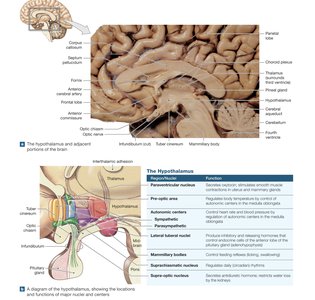
Nucleus | Function |
|---|---|
Paraventricular | Secretes oxytocin |
Supra-optic | Secretes ADH |
Autonomic centers | Control heart rate, BP, digestion |
Mammillary bodies | Relay olfactory info, control eating reflexes |
Suprachiasmatic | Regulates circadian rhythms |
Cerebrum Structure and Function
Cerebral Cortex and Hemispheres
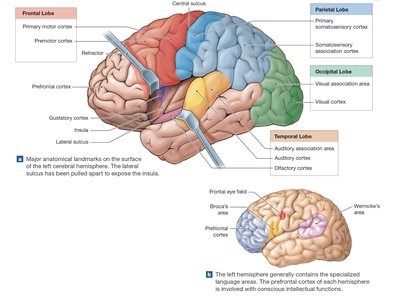
The cerebrum is divided into lobes (frontal, parietal, occipital, temporal, insula) and contains gyri and sulci to increase surface area. The cortex is responsible for conscious thought, sensory perception, and voluntary motor control.
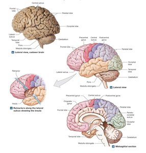
Lobe/Area | Function |
|---|---|
Frontal lobe | Voluntary motor control |
Parietal lobe | Somatosensory perception |
Occipital lobe | Visual perception |
Temporal lobe | Auditory and olfactory perception |
All lobes | Integration and processing |
White Matter of the Cerebrum
White matter consists of association, commissural, and projection fibers, connecting different regions within and between hemispheres, and linking the cortex to other brain regions.
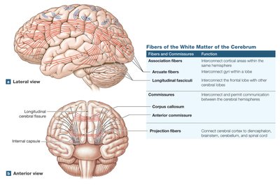
Fiber Type | Function |
|---|---|
Association fibers | Connect areas within one hemisphere |
Commissural fibers | Connect hemispheres (corpus callosum, anterior commissure) |
Projection fibers | Connect cortex to diencephalon, brainstem, cerebellum, spinal cord |
Basal Nuclei
Basal nuclei are deep masses of gray matter involved in subconscious control of skeletal muscle tone and coordination of learned movement patterns.
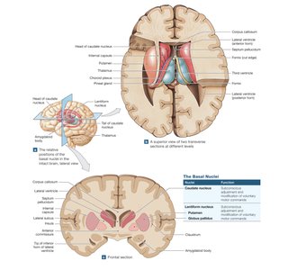
Nucleus | Function |
|---|---|
Caudate nucleus | Pattern and rhythm of movements |
Lentiform nucleus | Muscle tone and body position |
Putamen | Motor control |
Globus pallidus | Motor control |
Functional Areas of the Cortex
The cortex contains motor, sensory, and association areas. Motor areas control voluntary movements; sensory areas receive information from the body; association areas interpret and integrate data.
Electroencephalogram (EEG) and Brain Waves
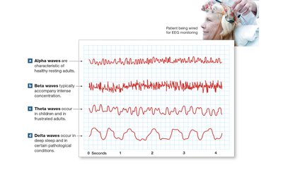
EEG records electrical activity of the brain, revealing patterns called brain waves. Types include alpha (resting), beta (concentration), theta (children/frustration), and delta (deep sleep/pathology).
Cranial Nerves
Overview and Classification
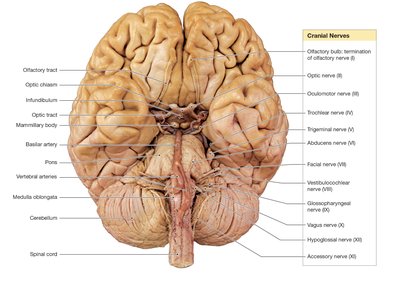
There are 12 pairs of cranial nerves, each with specific sensory, motor, or mixed functions. They are numbered I-XII based on their position along the brain's longitudinal axis.
Nerve | Primary Function |
|---|---|
I (Olfactory) | Smell |
II (Optic) | Vision |
III (Oculomotor) | Eye movement |
IV (Trochlear) | Eye movement |
V (Trigeminal) | Sensory/motor to face |
VI (Abducens) | Eye movement |
VII (Facial) | Sensory/motor to face |
VIII (Vestibulocochlear) | Balance/hearing |
IX (Glossopharyngeal) | Sensory/motor to head/neck |
X (Vagus) | Sensory/motor to thorax/abdomen |
XI (Accessory) | Motor to neck/back |
XII (Hypoglossal) | Tongue movement |
Cranial Reflexes
Somatic and Visceral Reflexes
Cranial reflexes are rapid, automatic responses involving cranial nerves. They are clinically important for assessing nerve function and include both somatic (skeletal muscle) and visceral (autonomic) responses.
Somatic reflexes: Simple, often used for clinical testing.
Visceral reflexes: Control autonomic functions (respiratory, digestive, cardiovascular).
Summary Table: Major Brain Regions and Functions
Region | Function |
|---|---|
Cerebrum | Conscious thought, memory, intellect, voluntary movement |
Cerebellum | Coordination of movement, balance |
Diencephalon | Sensory integration, autonomic control, hormone production |
Brainstem | Relay information, regulate vital functions |
Key Equations and Concepts
CSF Production:
EEG Frequency Ranges:
Alpha:
Beta:
Theta:
Delta:
Additional info:
Clinical conditions such as hydrocephalus, stroke, Bell's palsy, and Parkinson's disease are directly related to the anatomy and physiology of the brain and cranial nerves.
Mnemonic for cranial nerves: "Oh Oh Oh, To Touch And Feel Very Green Vegetables, Ah Heaven!"
