 Back
Study Guide: The Brain and Cranial Nerves (Chapter 14)
Study Guide - Smart Notes
 Tailored notes based on your materials, expanded with key definitions, examples, and context.
The Brain and Cranial Nerves
Introduction to the Brain and Cranial Nerves
The adult human brain is the central organ of the nervous system, containing nearly 97% of the body's nervous tissue. It weighs approximately 1.4 kg and has a volume of about 1200 mL. Despite differences in size between males and females, there is no correlation between brain size and intelligence.
Regions of the Brain
Main Brain Regions
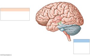
The brain is divided into four primary regions, each with distinct functions:
Cerebrum: Responsible for conscious thought, intellect, memory, and voluntary motor functions.
Cerebellum: Coordinates complex somatic motor patterns and adjusts output of other motor centers.
Diencephalon: Includes the thalamus (sensory relay) and hypothalamus (emotions, autonomic functions, hormone production).
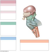
Brainstem: Relays information between the spinal cord and higher brain regions; includes the midbrain, pons, and medulla oblongata.
Brain Protection and Support
Physical and Biochemical Protection
The brain is protected by several structures:
Bones of the cranium: Provide rigid protection.
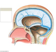
Cranial meninges: Three layers (dura mater, arachnoid mater, pia mater) that cover and protect the brain.
Cerebrospinal fluid (CSF): Cushions the brain, supports neural structures, and transports nutrients and waste.
Blood-brain barrier: Biochemical isolation that restricts passage of substances from blood to brain tissue.
Cerebrospinal Fluid (CSF) Circulation
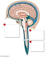
CSF is produced by the choroid plexus and circulates through the ventricles, central canal, and subarachnoid space, providing cushioning and nutrient transport.
The Brainstem
Medulla Oblongata
The medulla oblongata is the most inferior part of the brainstem, connecting the brain to the spinal cord. It regulates autonomic functions such as heart rate, blood pressure, and digestion, and contains nuclei for cranial nerves VIII-XII.
Reflex centers: Cardiovascular and respiratory rhythmicity centers.
Relay stations: Gracile and cuneate nuclei, solitary nuclei, and inferior olivary complex.
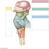
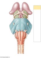
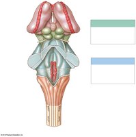
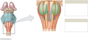
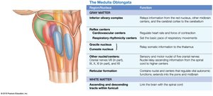
Functional Table: Medulla Oblongata
Region/Nucleus | Function |
|---|---|
Inferior olivary complex | Relays information to cerebellum about somatic motor commands |
Reflex centers | Regulate heart rate, blood flow, and respiratory rhythm |
Gracile and cuneate nuclei | Relay somatic sensory information to thalamus |
Other nuclei/centers | Sensory and motor nuclei of cranial nerves VIII-XII |
Reticular formation | Contains nuclei and centers regulating autonomic functions |
White matter | Ascending and descending tracts link brain with spinal cord |
Pons
The pons contains nuclei for cranial nerves, respiratory centers (apneustic and pneumotaxic), and tracts that relay information to and from the cerebellum.
Transverse pontine fibers: Link pons with cerebellar hemispheres.
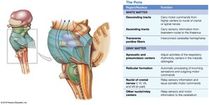
Functional Table: Pons
Region/Nucleus | Function |
|---|---|
Descending tracts | Carry motor commands from higher centers |
Ascending tracts | Carry sensory information |
Transverse fibers | Link pons with cerebellum |
Apneustic and pneumotaxic centers | Adjust rate and depth of respiration |
Reticular formation | Automatic processing of incoming sensations |
Nuclei of cranial nerves | Relay sensory and motor information |
Midbrain
The midbrain contains sensory nuclei (corpora quadrigemina), the red nucleus, substantia nigra, and cerebral peduncles. It processes visual and auditory information and maintains consciousness.
Superior colliculi: Visual information
Inferior colliculi: Auditory information
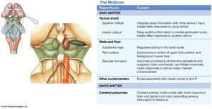
Functional Table: Midbrain
Region/Nucleus | Function |
|---|---|
Tectum (superior/inferior colliculi) | Integrates visual/auditory information, initiates reflexes |
Walls and floor (red nucleus, substantia nigra) | Regulate activity and muscle tone |
Reticular formation | Automatic processing of incoming sensations |
Other nuclei/centers | Nuclei associated with cranial nerves III and IV |
Cerebral peduncles | Connect primary motor cortex with motor neurons in brain and spinal cord |
The Cerebellum
Structure and Function
The cerebellum coordinates voluntary and involuntary movements, adjusts postural muscles, and fine-tunes motor activity. It contains the Purkinje cell layer and arbor vitae (white matter).
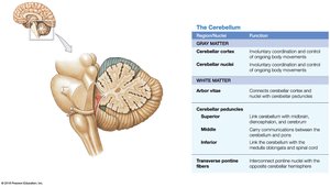
Functional Table: Cerebellum
Region/Nucleus | Function |
|---|---|
Cerebellar cortex | Involuntary coordination and control of movement |
Cerebellar nuclei | Relay information to Purkinje cells |
Arbor vitae | Connects cortex and nuclei |
Cerebellar peduncles | Link cerebellum with other brain regions |
Transverse pontine fibers | Connect pons nuclei with cerebellar hemispheres |
The Diencephalon
Thalamus and Hypothalamus
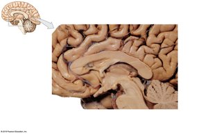
The diencephalon integrates sensory information and motor commands. The thalamus filters and relays sensory information, while the hypothalamus controls emotions, autonomic functions, and hormone production.
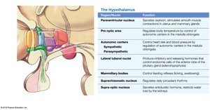
Functional Table: Hypothalamus
Region/Nucleus | Function |
|---|---|
Paraventricular nucleus | Secretes oxytocin, stimulates smooth muscle contraction |
Pre-optic area | Regulates body temperature |
Autonomic centers | Control heart rate and blood pressure |
Lateral tuberal nuclei | Produce inhibitory and releasing hormones |
Mammillary bodies | Control feeding reflexes |
Supra-optic nucleus | Secretes antidiuretic hormone (ADH) |
The Limbic System
Structure and Function
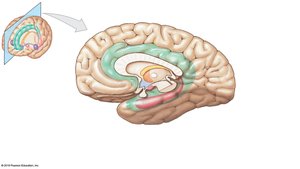
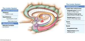
The limbic system establishes emotional states, links conscious and autonomic functions, and facilitates memory storage and retrieval. It includes the limbic lobe, cingulate gyrus, hippocampus, amygdaloid body, fornix, and thalamic nuclei.
Functional Table: Limbic System
Component | Function |
|---|---|
Limbic lobe cortical areas | Emotional processing |
Cingulate gyrus | Emotion and behavior regulation |
Hippocampus | Memory formation |
Amygdaloid body | Emotional responses |
Reticular formation | Alertness and sleep regulation |
The Cerebrum
Structure and Functional Areas
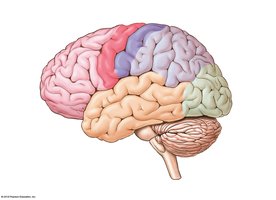
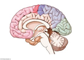
The cerebrum is the largest part of the brain, responsible for conscious thought, intellectual functions, and processing somatic sensory and motor information. It contains gyri, sulci, and fissures that increase surface area for cortical neurons.
White Matter of the Cerebrum
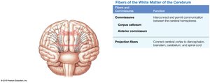
White matter consists of association fibers (within one hemisphere), commissural fibers (between hemispheres), and projection fibers (link cortex to other brain regions).
Functional Table: White Matter Fibers
Fiber Type | Function |
|---|---|
Commissures | Permit communication between hemispheres |
Corpus callosum | Main commissural fiber bundle |
Anterior commissure | Connects temporal lobes |
Projection fibers | Connect cortex to diencephalon, brainstem, cerebellum, and spinal cord |
Functional Areas of the Cerebral Cortex
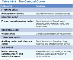
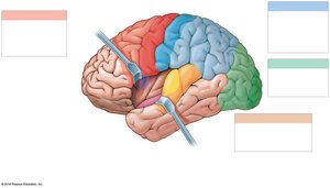
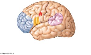
The cerebral cortex is divided into lobes, each with specialized functions:
Lobe/Area | Function |
|---|---|
Frontal lobe | Primary motor cortex: voluntary control of skeletal muscles |
Parietal lobe | Primary somatosensory cortex: conscious perception of touch, pressure, pain, vibration, temperature |
Occipital lobe | Visual cortex: conscious perception of visual stimuli |
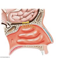
Temporal lobe | Auditory and olfactory cortices: perception of hearing and smell |
All lobes | Integration and processing of sensory data; initiation of motor activities |
Motor, Sensory, and Association Areas
Motor areas (precentral gyrus), sensory areas (postcentral gyrus), and association areas (premotor cortex, somatosensory association cortex, visual and auditory association areas) interpret and coordinate information.
Cranial Nerves
Overview and Classification
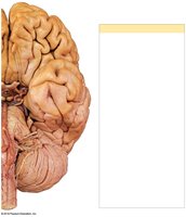
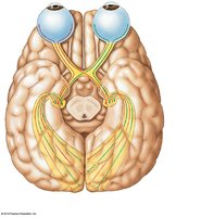
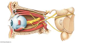
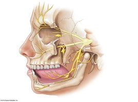
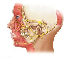
There are 12 pairs of cranial nerves, classified as sensory, motor, mixed, or special sensory. Each nerve has specific functions related to sensation, movement, or autonomic control.
Selected Cranial Nerves and Functions
Olfactory nerve (I): Special sensory (smell)
Optic nerve (II): Special sensory (vision)
Oculomotor nerve (III): Eye movements
Trochlear nerve (IV): Eye movement (superior oblique)
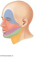
Trigeminal nerve (V): Sensory and motor of face
Abducens nerve (VI): Eye movements (lateral rectus)
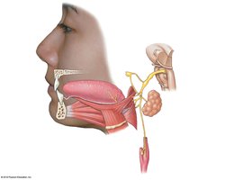
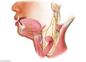
Facial nerve (VII): Sensory and motor of face
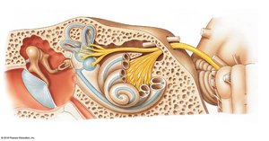
Vestibulocochlear nerve (VIII): Balance and hearing
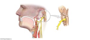
Glossopharyngeal nerve (IX): Sensory and motor of head and neck
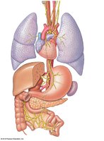
Vagus nerve (X): Sensory and motor of parasympathetic nervous system
Accessory nerve (XI): Muscles of neck and upper back
Hypoglossal nerve (XII): Tongue movements
Cranial Reflexes
Cranial reflexes involve sensory and motor fibers of cranial nerves and are clinically useful for assessing nerve and brain function.
Summary Table: Major Brain Regions and Functions
Region | Function |
|---|---|
Cerebrum | Conscious thought, intellect, memory, voluntary motor control |
Cerebellum | Coordination of movement, balance, fine-tuning motor activity |
Diencephalon | Sensory relay, autonomic and endocrine control |
Brainstem | Relay information, regulate autonomic functions, cranial nerve nuclei |
Additional info: This study guide expands on brief points from the original materials, providing definitions, examples, and tables for clarity. All included images are directly relevant to the adjacent content and reinforce anatomical and functional concepts.
