 Back
Study Guide: The Cardiovascular System – Blood Vessels
Study Guide - Smart Notes
 Tailored notes based on your materials, expanded with key definitions, examples, and context.
The Cardiovascular System: Blood Vessels
Overview of Blood Vessels
The cardiovascular system consists of a network of blood vessels that transport blood throughout the body. These vessels are classified as arteries, capillaries, and veins, each serving distinct functions in circulation.
Arteries: Carry blood away from the heart; most are oxygenated except for pulmonary arteries and fetal umbilical vessels.
Capillaries: Microscopic vessels that directly serve tissue needs by facilitating exchange of gases, nutrients, and wastes.
Veins: Return blood to the heart; most are deoxygenated except for pulmonary veins.
Example: The aorta is the largest artery, while the vena cava is the largest vein.
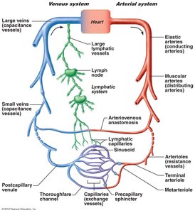
Structure of Blood Vessel Walls
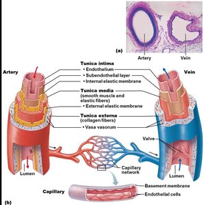
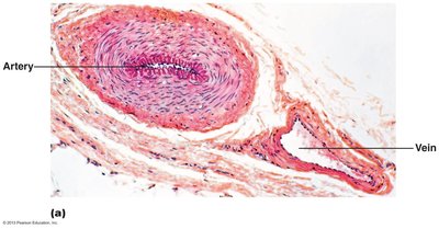
Blood vessel walls are composed of three layers (tunics) in arteries and veins, while capillaries have a single layer.
Tunica intima: Innermost layer; consists of endothelium and, in larger vessels, a subendothelial layer.
Tunica media: Middle layer; contains smooth muscle and elastic fibers, responsible for vasoconstriction and vasodilation.
Tunica externa (adventitia): Outermost layer; composed of collagen fibers, provides structural support and protection.
Lumen: Central space through which blood flows.
Capillaries: Composed only of endothelium and a sparse basal lamina.
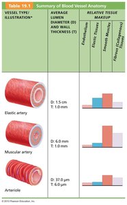
Types of Arteries
Arteries are classified based on their size and function.
Elastic (Conducting) Arteries: Largest arteries (e.g., aorta); contain abundant elastin, act as pressure reservoirs.
Muscular (Distributing) Arteries: Medium-sized; deliver blood to organs, have more smooth muscle, active in vasoconstriction.
Arterioles: Smallest arteries; regulate blood flow into capillary beds via vasodilation and vasoconstriction.
Vessel Type | Diameter (D) | Wall Thickness (T) | Relative Tissue Makeup |
|---|---|---|---|
Elastic artery | 1.5 cm | 1.0 mm | High elastin, moderate smooth muscle, moderate fibrous tissue |
Muscular artery | 0.4 cm | 1.0 mm | Moderate elastin, high smooth muscle, moderate fibrous tissue |
Arteriole | 37.0 μm | 6.0 μm | Low elastin, moderate smooth muscle, low fibrous tissue |
Capillaries: Structure and Function
Capillaries are the smallest blood vessels and are essential for exchange between blood and tissues. Their walls are one cell thick, allowing only a single red blood cell to pass at a time.
Pericytes: Contractile cells that stabilize capillary walls and regulate permeability.
Functions: Exchange of gases, nutrients, wastes, and hormones.
Distribution: Found in all tissues except cartilage, epithelia, cornea, and lens.
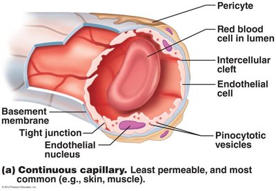
Types of Capillaries
Capillaries are classified based on their permeability and structure.
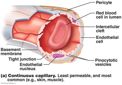
Continuous Capillaries: Least permeable, most common; found in skin and muscles. Tight junctions connect endothelial cells, but intercellular clefts allow passage of fluids and small solutes. In the brain, tight junctions are complete, forming the blood-brain barrier.
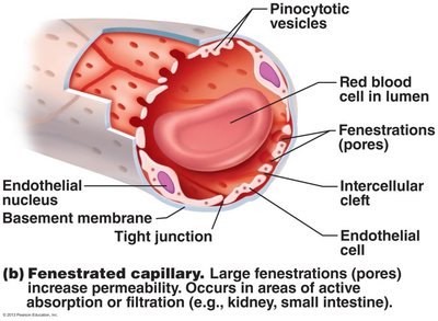
Fenestrated Capillaries: Have pores (fenestrations) that increase permeability; found in areas of active absorption or filtration (e.g., kidneys, small intestine).
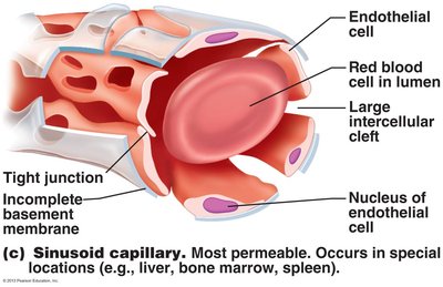
Sinusoidal Capillaries (Sinusoids): Most permeable; have large intercellular clefts and incomplete basement membranes. Found in liver, bone marrow, and spleen.
Capillary Beds and Microcirculation
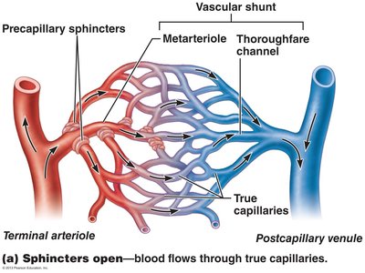
Capillary beds are networks of capillaries that connect arterioles and venules, facilitating microcirculation. Blood flow through capillary beds is regulated by precapillary sphincters.
Vascular shunt: Direct connection between terminal arteriole and postcapillary venule.
True capillaries: Branch off the metarteriole; 10–100 exchange vessels per capillary bed.
Precapillary sphincters: Regulate blood flow into true capillaries based on local chemical conditions and neural input.
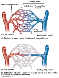
Venous System: Venules and Veins
Venules are formed when capillary beds unite and are very porous, allowing fluids and white blood cells to enter tissues. Veins are formed when venules converge and serve as blood reservoirs.
Veins: Have thinner walls and larger lumens than arteries; contain up to 65% of the blood supply.
Adaptations for venous return: Large-diameter lumens, valves to prevent backflow, and the milking effect of skeletal muscles.
Venous sinuses: Flattened veins with extremely thin walls (e.g., coronary sinus, dural sinuses).
Vascular Anastomoses
Anastomoses are interconnections between blood vessels that provide alternate pathways for blood flow.
Arterial anastomoses: Common at joints, abdominal organs, brain, and heart; provide collateral circulation.
Venous anastomoses: More common; blockage rarely leads to tissue death.
Arteriovenous anastomoses: Vascular shunts in capillaries.
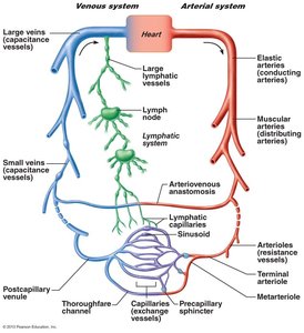
Physiology of Circulation
Key Terms
Blood flow (F): Volume of blood flowing through a vessel, organ, or entire circulation per unit time (ml/min).
Blood pressure (BP): Force per unit area exerted on vessel wall by blood; measured in mm Hg.
Resistance (R): Opposition to blood flow; determined by blood viscosity, vessel length, and vessel diameter.
Relationship Between Blood Flow, Pressure, and Resistance
Blood flow (F) is directly proportional to the pressure gradient (ΔP):
Resistance is inversely proportional to vessel radius:
Systemic blood pressure: Highest in the aorta, declines throughout the pathway, steepest drop in arterioles.
Arterial Blood Pressure
Systolic pressure: Pressure during ventricular contraction.
Diastolic pressure: Lowest arterial pressure during relaxation.
Pulse pressure: Difference between systolic and diastolic pressure.
Mean arterial pressure (MAP): Average pressure propelling blood to tissues. Formula:
Example Calculation
If systolic = 120 mmHg, diastolic = 80 mmHg:
Capillary and Venous Blood Pressure
Capillary blood pressure: Ranges from 35 to 15 mm Hg; low pressure prevents rupture and allows exchange.
Venous blood pressure: Small gradient (~15 mm Hg); aided by respiratory and muscular pumps, and venous vasoconstriction.
Maintaining Blood Pressure
Blood pressure is regulated by the heart, blood vessels, kidneys, and brain. The main factors influencing BP are cardiac output (CO), peripheral resistance (PR), and blood volume.
Relationship:
Changes in one variable are compensated by changes in others to maintain BP.
Control of Blood Pressure
Short-term regulation: Neural and hormonal controls alter peripheral resistance and cardiac output.
Long-term regulation: Renal mechanisms alter blood volume.
Neural Controls: Baroreceptor and Chemoreceptor Reflexes
Baroreceptors: Located in carotid sinuses, aortic arch, and large arteries; respond to changes in pressure.
Vasomotor center: Cluster of sympathetic neurons in the medulla; regulates vessel diameter.
Chemoreceptors: Respond to changes in CO2, pH, and O2; increase BP and respiratory rate.
Hormonal Controls
Adrenal medulla hormones: Norepinephrine and epinephrine increase cardiac output and cause vasoconstriction.
Angiotensin II: Potent vasoconstrictor, increases BP.
Antidiuretic hormone (ADH): Causes vasoconstriction in cases of low BP.
Atrial natriuretic peptide (ANP): Causes vasodilation and decreases blood volume and BP.
Renal Regulation of Blood Pressure
Direct mechanism: Kidneys eliminate or conserve water to regulate blood volume and BP.
Indirect mechanism: Renin-angiotensin system; renin release leads to angiotensin II production, causing vasoconstriction and aldosterone secretion.
Monitoring Circulatory Efficiency
Vital Signs and Blood Pressure Measurement
Pulse: Pressure wave caused by expansion and recoil of arteries; radial pulse is commonly measured.
Blood pressure: Measured indirectly using a sphygmomanometer and auscultatory method.
Systolic pressure: First sound heard as blood spurts through the artery.
Diastolic pressure: Pressure when sounds disappear.
Variations and Alterations in Blood Pressure
Hypotension: Low blood pressure; may be associated with long life or circulatory shock.
Hypertension: High blood pressure; major risk factor for heart failure, stroke, and renal disease.
Primary hypertension: No identifiable cause; associated with risk factors like heredity, diet, obesity, and stress.
Secondary hypertension: Due to identifiable disorders (e.g., kidney disease, endocrine disorders).
Blood Flow Through Body Tissues
Tissue Perfusion and Autoregulation
Tissue perfusion: Delivery of O2 and nutrients, removal of wastes, gas exchange, absorption, and urine formation.
Autoregulation: Automatic adjustment of blood flow to meet tissue needs; controlled by local arterioles.
Metabolic controls: Vasodilation in response to low O2, high CO2, and other metabolites.
Myogenic controls: Vascular smooth muscle responds to changes in pressure to maintain constant perfusion.
Angiogenesis: Formation of new blood vessels when short-term regulation is insufficient.
Blood Flow in Specific Organs
Skeletal muscles: Blood flow increases with activity; local controls override sympathetic vasoconstriction.
Brain: Constant blood flow; sensitive to pH and CO2 changes.
Skin: Blood flow regulated for nutrient supply, temperature control, and as a reservoir.
Lungs: Low pressure circuit; vasoconstriction occurs in response to low O2.
Heart: Blood flow increases during exercise; coronary vessels dilate in response to local vasodilators.
Capillary Exchange and Fluid Movements
Vasomotion: Intermittent flow through capillaries due to opening/closing of precapillary sphincters.
Exchange mechanisms: Diffusion (O2, CO2, nutrients, wastes), passage through clefts/fenestrations, and active transport.
Bulk flow: Movement of fluid determined by hydrostatic and osmotic pressures.
Hydrostatic pressure: Forces fluid out of capillaries; higher at arterial end.
Colloid osmotic pressure: Draws fluid into capillaries; created by plasma proteins.
Net filtration pressure (NFP):
Excess fluid is returned to the blood via the lymphatic system.
