 Back
BackStudy Guide: The Cardiovascular System – Blood Vessels and Circulation
Study Guide - Smart Notes
 Tailored notes based on your materials, expanded with key definitions, examples, and context.
The Cardiovascular System: Blood Vessels and Circulation
Introduction to Blood Vessels
The cardiovascular system is composed of a network of blood vessels that transport blood throughout the body. These vessels are classified into three main types: arteries, veins, and capillaries. Each type plays a distinct role in circulation:
Arteries: Carry blood away from the heart.
Veins: Return blood toward the heart.
Capillaries: Connect arteries and veins, facilitating exchange of substances between blood and tissues.
Histological Organization of Blood Vessels
Blood vessel walls are organized into three layers, each with specific structural and functional properties:
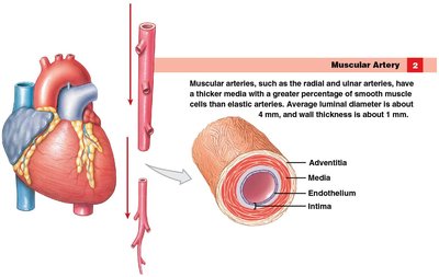
Adventitia (Tunica Externa): Outermost layer, composed of connective tissue (collagen and elastic fibers) for support and anchoring.
Media (Tunica Media): Middle layer, primarily smooth muscle, responsible for regulating vessel diameter and blood flow.
Intima (Tunica Intima): Innermost layer, a simple squamous epithelium (endothelium) that lines the lumen.
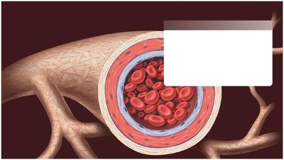
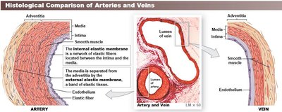
Arteries and veins possess all three layers, while capillaries consist only of the intima, allowing efficient exchange of substances.
Structural Differences Between Arteries and Veins
Arteries and veins differ in wall thickness, elasticity, and lumen size:
Arteries: Thicker media, maintain shape, extra elastic membranes, smaller lumen.
Veins: Thinner media, often thicker adventitia, larger lumen, can hold more blood, may collapse in cross-section.
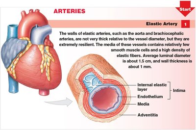
Types of Arteries
Arteries are classified by size and function:
Elastic Arteries: Largest arteries (e.g., aorta), thick elastic membranes, expand and recoil to maintain blood flow.
Muscular Arteries: Medium-sized (e.g., carotid, brachial), thick media, regulate blood flow to organs.
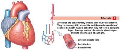
Arterioles: Smallest arteries, thin adventitia, control blood flow into capillaries.
Types of Capillaries
Capillaries are the smallest blood vessels, specialized for exchange:
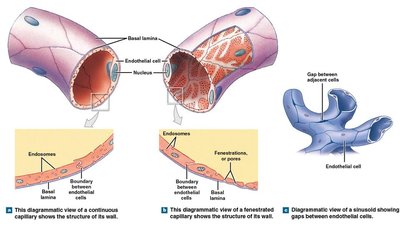
Continuous Capillaries: Complete endothelium, most common, allow passage of small molecules and gases.
Fenestrated Capillaries: Endothelium with pores (fenestrations), facilitate rapid transport and passage of larger molecules.
Sinusoids (Sinusoidal Capillaries): Large gaps in endothelium, irregular shape, allow movement of proteins and cells (e.g., in liver, bone marrow).
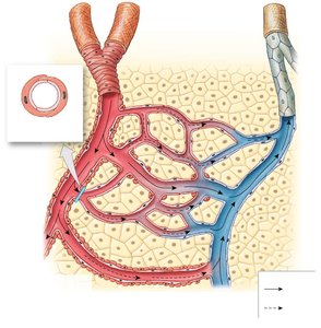
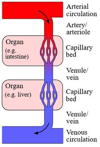
Capillary Beds
Capillaries form networks called capillary beds that supply tissues with oxygen and nutrients. Capillary beds provide multiple pathways for blood flow, ensuring tissue perfusion even if some vessels are blocked.
Multiple arterioles can supply the same capillary bed.
Blood flow through capillary beds is regulated by precapillary sphincters.
Types of Veins
Veins are categorized by size and function:
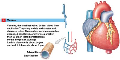
Venules: Smallest veins, collect blood from capillary beds, thin walls.
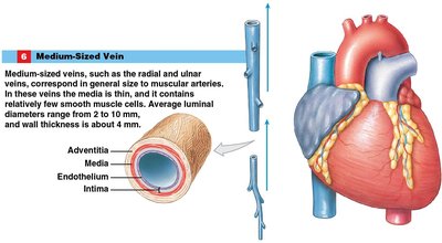
Medium-Sized Veins: Larger, contain valves to prevent backflow, thinner media, thicker adventitia.
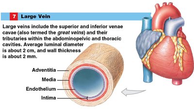
Large Veins: Largest veins (e.g., superior and inferior vena cava), thick adventitia, no valves.
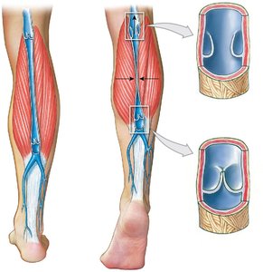
Venous Valves and Blood Flow
Medium-sized veins contain venous valves that prevent backflow, especially in the lower limbs. Skeletal muscle contractions help propel blood toward the heart.
Distribution of Blood in the Cardiovascular System
Blood is unevenly distributed between arteries and veins:
Veins contain about two-thirds of the body's blood volume.
Veins can expand to accommodate more blood (venous reservoirs).
Venoconstriction can shift blood to arteries during increased demand.
Parallel Arrangement and Naming of Blood Vessels
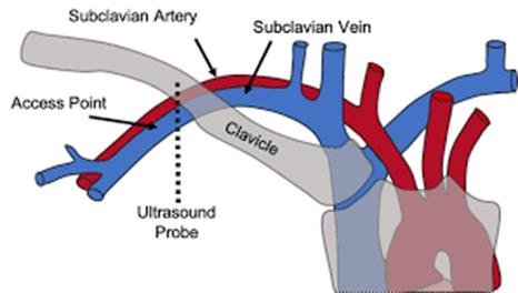
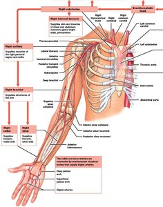
Many arteries and veins run parallel and share names (e.g., subclavian artery and vein). Most veins are deep, but some are superficial and used for IVs and blood draws.
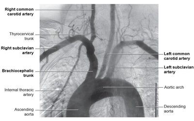
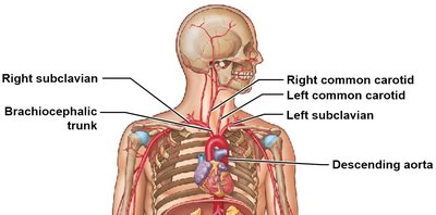
Major Blood Vessels: The Aorta and Its Branches
The aorta is the largest artery, divided into:
Ascending Aorta: Originates from the left ventricle, gives rise to coronary arteries.
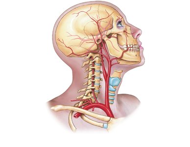
Aortic Arch: Curves and branches into major arteries for head, neck, and arms.
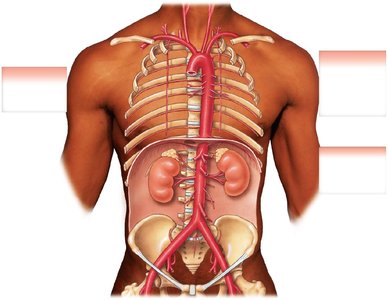
Descending Aorta: Travels through thoracic and abdominal cavities, supplies organs and lower limbs.
Systemic Veins and Portal Systems
Veins return blood to the heart, often paralleling arteries. The jugular veins return blood from the head and neck. Blood from above the diaphragm enters via the superior vena cava, and from below via the inferior vena cava.
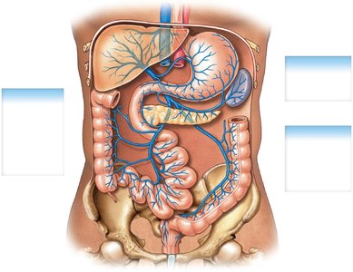
Some vessels form portal systems, carrying blood through multiple capillary beds before returning to the heart. The hepatic portal system delivers blood from the digestive tract to the liver for detoxification and nutrient processing.
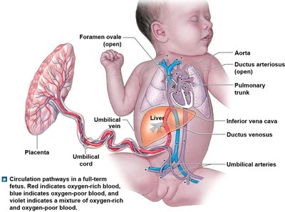
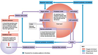
Cardiovascular Changes at Birth
The fetal cardiovascular system differs from the adult system:
Oxygen and nutrients are received from the mother via the placenta.
Umbilical arteries: Carry deoxygenated blood to the placenta.
Umbilical vein: Returns oxygenated blood to the fetus, bypassing the liver via the ductus venosus.
Foramen ovale: Opening between right and left atria, allows blood to bypass lungs.
Ductus arteriosus: Connects pulmonary trunk to aortic arch, further bypassing lungs.
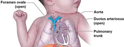
Transition at Birth
At birth, the first breath causes the lungs to expand, blood flow through the pulmonary circuit increases, and fetal shunts close:
Ductus arteriosus: Constricts and closes, forcing blood through the lungs.
Foramen ovale: Closes due to increased left atrial pressure, becoming the fossa ovalis.
Example: The closure of fetal shunts is essential for normal postnatal circulation and oxygenation. Additional info: These changes are critical for the transition from placental to independent pulmonary respiration.
