 Back
Study Guide: The Cardiovascular System – Heart and Blood Vessels
Study Guide - Smart Notes
 Tailored notes based on your materials, expanded with key definitions, examples, and context.
The Cardiovascular System
Overview and Functions
The cardiovascular system consists of the heart, blood, and blood vessels. It is responsible for transporting blood throughout the body, delivering essential substances, removing waste, distributing hormones, and regulating body temperature.
Arteries: Carry blood away from the heart; serve as distribution channels to organs.
Veins: Return blood to the heart; serve as blood reservoirs.
Capillaries: Small vessels connecting arteries and veins; permit diffusion of gases, nutrients, and waste.
The system is subdivided into:
Pulmonary Circuit: Transports blood between the heart and lungs for gas exchange.
Systemic Circuit: Transports blood between the heart and the rest of the body.
Functions:
Transportation of oxygen, nutrients, and hormones
Removal of metabolic byproducts (CO2, waste)
Thermoregulation
Anatomy of the Heart
General Characteristics
The heart is a muscular organ, about the size of a fist, located within the mediastinum of the thoracic cavity. It is positioned anterior to the vertebral column and posterior to the sternum, resting on the diaphragm.
Base: Superior portion, attached to great vessels.
Apex: Inferior, pointed tip (left ventricle); point of maximal intensity (PMI).
Borders: Superior (great vessels), Inferior (ventricles), Right (right atrium), Left (left ventricle).
Coverings of the Heart
Pericardium: Double-walled sac enclosing the heart.
Fibrous Pericardium: Tough, dense connective tissue; protects, anchors, prevents overfilling.
Serous Pericardium: Two layers (parietal and visceral/epicardium); contains pericardial cavity with lubricating fluid.
Clinical Conditions:
Pericarditis: Inflammation of pericardium; causes pain and friction rub.
Cardiac Tamponade: Compression due to fluid accumulation; prevents proper ventricular filling.
Layers of the Heart Wall
Epicardium: Visceral pericardium; outermost layer.
Myocardium: Middle layer; cardiac muscle tissue; contracts to pump blood.
Endocardium: Innermost layer; simple squamous epithelium; lines chambers and continuous with vessel endothelium.
Chambers of the Heart
Four chambers: 2 atria (upper), 2 ventricles (lower).
Interatrial and Interventricular Septa: Divide chambers longitudinally.
Coronary Sulcus: Marks border between atria and ventricles; contains coronary vessels.
Auricle: Flap on atria; right auricle is larger.
Atria
Receiving chambers for blood returning from systemic (right atrium) and pulmonary (left atrium) circuits.
Thin-walled; contain pectinate muscles and fossa ovalis (remnant of foramen ovale).
Right atrium receives blood via superior vena cava, inferior vena cava, and coronary sinus.
Left atrium receives blood via four pulmonary veins (oxygenated).
Ventricles
Thick-walled; pump blood to pulmonary (right ventricle) and systemic (left ventricle) circuits.
Contain trabeculae carneae and papillary muscles.
Right ventricle pumps blood through pulmonary semilunar valve to pulmonary trunk.
Left ventricle pumps blood through aortic semilunar valve to aorta.
Heart Valves
Valves ensure unidirectional blood flow:
Atrioventricular (AV) Valves: Tricuspid (right), Bicuspid/Mitral (left); prevent backflow into atria.
Semilunar Valves: Pulmonary (right), Aortic (left); prevent backflow into ventricles.
Chordae Tendineae: Collagen cords anchoring AV valve flaps to papillary muscles.
Valvular Disease:
Regurgitation/Prolapse: Valve does not close properly; blood leaks backward.
Stenosis: Valve does not open fully; increases workload.
The Cardiac Circuit
Pulmonary, Systemic, and Coronary Circuits
Pulmonary Circuit: Right heart pumps deoxygenated blood to lungs.
Systemic Circuit: Left heart pumps oxygenated blood to body.
Coronary Circuit: Supplies heart muscle via coronary arteries and veins.
Coronary Circulation
Right Coronary Artery: Marginal and posterior interventricular arteries.
Left Coronary Artery: Anterior interventricular and circumflex arteries.
Cardiac Veins: Great, middle, small, posterior, and anterior cardiac veins; drain into coronary sinus.
Pathway of Blood Through the Heart
Deoxygenated blood: SVC/IVC/coronary sinus → right atrium → tricuspid valve → right ventricle → pulmonary semilunar valve → pulmonary trunk → pulmonary arteries → lungs
Oxygenated blood: pulmonary veins → left atrium → bicuspid valve → left ventricle → aortic semilunar valve → aorta → body/coronary arteries
Cardiac Muscle Cells
Types and Structure
Autorhythmic (Conducting) Cells: 1%; spontaneously depolarize; set heart rate.
Contractile Cells: 99%; produce force for pumping; striated, branched, involuntary.
Intercalated Discs: Connect cells; contain gap junctions (electrical) and desmosomes (physical).
Physiology of the Heart
The Conducting System
Sinoatrial Node (SA): Pacemaker; fastest depolarization; initiates heartbeat.
Atrioventricular Node (AV): Delays impulse; allows atrial contraction.
AV Bundle (Bundle of His): Only electrical connection between atria and ventricles.
Bundle Branches: Carry impulse to apex.
Purkinje Fibers: Distribute impulse to ventricles.
Intrinsic and Extrinsic Control
Intrinsic: Autorhythmic cells set rate.
Extrinsic: Medulla oblongata (cardioacceleratory and cardioinhibitory centers), hormones (epinephrine, thyroxine).
Cardiac Action Potential
Slow Fibers: SA and AV nodes; slow depolarization.
Fast Fibers: Atrial/ventricular muscle, Bundle of His, Purkinje fibers; rapid depolarization.
Phases:
Phase 0: Na+ influx, rapid depolarization
Phase 1: Peak, Na+ channels close, K+ exits
Phase 2: Plateau, Ca2+ influx
Phase 3: Repolarization, K+ exits
Phase 4: Return to resting potential
Mechanisms of Contraction
Na+ influx → depolarization → Ca2+ release from SR and extracellular space → binds troponin → contraction
Cardiac muscle requires Ca2+ from both SR and extracellular fluid.
Differences Between Cardiac and Skeletal Muscle
Stimulation: Cardiac is self-excitable; skeletal requires nerve.
Refractory Period: Cardiac has longer ARP; prevents tetanus.
Cardiodynamics
Cardiac Cycle
The cardiac cycle includes all events associated with blood flow during one heartbeat, including contraction (systole) and relaxation (diastole) of all four chambers. Blood moves from areas of higher to lower pressure.
Ventricular Filling: Passive filling (80%), then atrial contraction (20%) – end-diastolic volume (EDV).
Ventricular Systole: Isovolumetric contraction (AV valves close, S1), then ventricular ejection (semilunar valves open, blood ejected, end-systolic volume (ESV)).
Isovolumetric Relaxation: Semilunar valves close (S2), ventricular pressure falls, AV valves open, cycle repeats.
Right vs. Left Ventricle: Both contract simultaneously with identical stroke volumes; left ventricle operates under higher pressure.
Heart Sounds
S1 (Lub): AV valves close during ventricular systole.
S2 (Dub): Semilunar valves close during ventricular diastole.
S3: Blood flowing into ventricles; faint.
S4: Atrial contraction; usually pathological.
Regulation of the Cardiac Cycle
SA Node: Primary controller of heart rate.
Parasympathetic: Decreases heart rate (ACh).
Sympathetic: Increases heart rate and contractility (NE).
Cardiac Control Center: Medulla oblongata; balances sympathetic and parasympathetic input.
Key Terms, Definitions & Units
Cardiac Output (CO): Blood pumped by each ventricle per minute.
Heart Rate (HR): Beats per minute.
Stroke Volume (SV): Blood pumped per beat.
EDV: End-diastolic volume; volume at end of diastole.
ESV: End-systolic volume; volume at end of systole.
Ejection Fraction: Percentage of EDV ejected per beat.
Blood Pressure: Force against arterial walls (mm Hg).
Systolic/Diastolic Pressure: Peak/lowest arterial pressure.
Blood Volume: Total blood in system (4-6 L).
Preload: Degree of ventricular stretch during diastole.
Afterload: Tension needed to open semilunar valves.
Cardiac Output and Stroke Volume
Formulas and Factors
Cardiac Output:
Stroke Volume:
Average CO: 5.4 L/min
Factors affecting SV: preload, afterload, contractility
Preload
Degree of stretch; directly proportional to EDV
Frank-Starling Law: Greater stretch → greater force of contraction
Afterload
Pressure to overcome in large vessels; increased afterload decreases SV
Contractility
Independent of stretch; increased Ca2+ influx increases contractility
Positive inotropic factors: digitalis, Ca2+, glucagon, thyroxine
Negative inotropic factors: excess H+, hyperkalemia, calcium channel blockers
Regulation of Heart Rate
Autonomic Nervous System: Sympathetic increases HR and contractility; parasympathetic decreases HR.
Baroreceptors: Monitor blood pressure; adjust HR and SV to maintain homeostasis.
Chemical Regulation: Hormones (epinephrine, thyroxine), ions (K+, Ca2+).
Other Factors: Age, gender, exercise.
Baroreceptor Reflex
Located in aortic arch and carotid sinuses; monitor blood pressure.
Increase in BP → increased cardioinhibitory activity → decreased HR and SV → decreased CO and BP.
Decrease in BP → increased cardioacceleratory activity → increased HR and SV → increased CO and BP.
Congestive Heart Failure (CHF)
Contractile dysfunction and hemodynamic insufficiency.
Left ventricular remodeling → dilation, fibrosis, contractile failure.
Left-sided failure → pulmonary edema; right-sided failure → systemic congestion.
Common causes: MI, ischemic cardiomyopathy, CAD.
Risk factors: hypertension, diabetes, hypercholesterolemia, cardiomyopathies.
Cardiac Cycle: Pressure-Volume Relationship
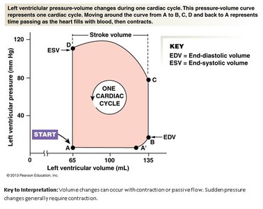
Pressure-Volume Loop
The pressure-volume loop illustrates changes in left ventricular pressure and volume during one cardiac cycle. Moving around the curve from A to B, C, D, and back to A represents time as the heart fills with blood, then contracts.
EDV (End-Diastolic Volume): Maximum volume at end of filling.
ESV (End-Systolic Volume): Minimum volume at end of contraction.
Stroke Volume: Difference between EDV and ESV.
Volume changes can occur with contraction or passive flow; sudden pressure changes generally require contraction.
Additional info: The pressure-volume loop is a fundamental tool for understanding cardiac mechanics and is used clinically to assess cardiac function and diagnose heart disease.
