 Back
BackStudy Guide: The Cardiovascular System – The Heart and Blood Vessels
Study Guide - Smart Notes
 Tailored notes based on your materials, expanded with key definitions, examples, and context.
The Heart: Structure and Function
Gross Anatomy of the Heart
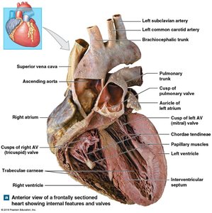
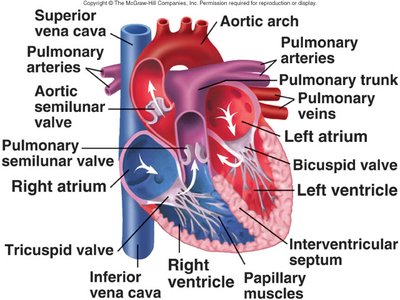
The heart is a muscular organ responsible for pumping blood throughout the body via the circulatory system. It consists of four chambers: two atria and two ventricles, separated by valves that ensure unidirectional blood flow.
Right Atrium: Receives deoxygenated blood from the body via the superior and inferior vena cava.
Right Ventricle: Pumps deoxygenated blood to the lungs through the pulmonary trunk.
Left Atrium: Receives oxygenated blood from the lungs via the pulmonary veins.
Left Ventricle: Pumps oxygenated blood to the body through the aorta.
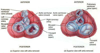
Valves: Tricuspid (right AV), bicuspid/mitral (left AV), pulmonary semilunar, and aortic semilunar valves prevent backflow.
Interventricular Septum: Separates the right and left ventricles.
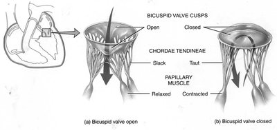
Chordae Tendineae and Papillary Muscles: Anchor AV valves and prevent prolapse during ventricular contraction.
Layers of the Heart Wall
The heart wall is composed of three layers:
Epicardium: The outer layer, consisting of connective tissue.
Myocardium: The middle layer, made of cardiac muscle tissue responsible for contraction.
Endocardium: The inner layer, consisting of endothelial cells lining the chambers.
Blood Flow Through the Heart
Pulmonary and Systemic Circulation
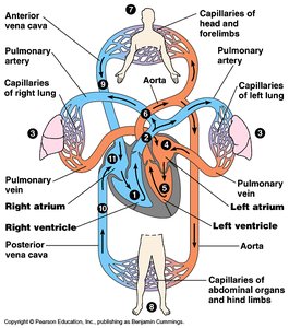
The heart acts as a dual pump, directing blood through two main circuits:
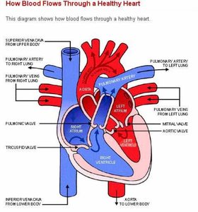
Pulmonary Circulation: Right ventricle → pulmonary arteries → lungs → pulmonary veins → left atrium.
Systemic Circulation: Left ventricle → aorta → body tissues → vena cavae → right atrium.
Valves and Blood Flow Regulation
Heart valves ensure unidirectional blood flow and prevent regurgitation:
AV Valves: Tricuspid (right) and bicuspid/mitral (left) valves separate atria from ventricles.
Semilunar Valves: Pulmonary and aortic valves separate ventricles from major arteries.
Valves open and close in response to pressure changes during the cardiac cycle.
The Cardiac Cycle
Phases of the Cardiac Cycle
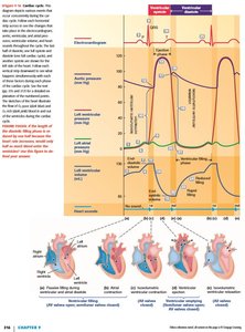
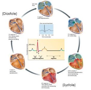
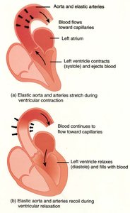
The cardiac cycle consists of alternating periods of contraction (systole) and relaxation (diastole):
Ventricular Filling: Blood flows from atria to ventricles (diastole).
Isovolumetric Contraction: Ventricles contract with all valves closed, building pressure.
Ventricular Ejection: Semilunar valves open, blood is pumped out.
Isovolumetric Relaxation: Ventricles relax, all valves closed, pressure drops.
Pressure Changes and Valve Function
Pressure changes in the heart chambers and vessels drive the opening and closing of valves:
During ventricular contraction, pressure rises, AV valves close, and semilunar valves open.
During relaxation, pressure falls, semilunar valves close, and AV valves open.
Electrical Activity of the Heart
Intrinsic Conduction System
The heart's electrical activity is coordinated by specialized autorhythmic cells:
SA Node: Pacemaker cells initiate action potentials.
AV Node: Delays impulse, allowing atrial contraction.
Bundle of His, Bundle Branches, Purkinje Fibers: Rapidly conduct impulses to ventricles.
ECG/EKG Interpretation
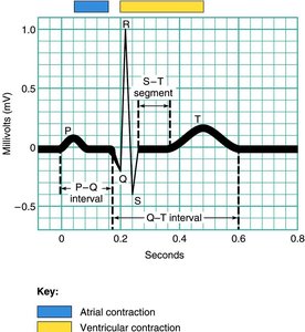
An electrocardiogram (ECG/EKG) records the heart's electrical events:
P Wave: Atrial depolarization.
QRS Complex: Ventricular depolarization.
T Wave: Ventricular repolarization.
Cardiac Muscle Physiology
Action Potentials in Cardiac Muscle
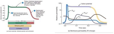
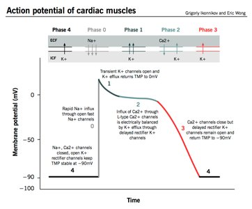
Cardiac muscle cells exhibit unique action potentials with a plateau phase:
Rapid Depolarization: Opening of voltage-gated Na+ channels.
Plateau Phase: Slow influx of Ca2+ through L-type channels maintains depolarization.
Repolarization: Closure of Ca2+ channels and opening of K+ channels.
The plateau prevents tetanus and allows sufficient time for blood ejection.
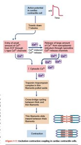
Excitation-Contraction Coupling
Calcium ions play a critical role in cardiac muscle contraction:
Ca2+ influx from extracellular fluid triggers further Ca2+ release from the sarcoplasmic reticulum (CICR).
Ca2+ binds to troponin, allowing actin-myosin crossbridge cycling.
Contraction strength depends on Ca2+ availability.
Relaxation occurs as Ca2+ is removed by pumps and exchangers.
Autorhythmic Cells and Pacemaker Activity
Action Potentials in Autorhythmic Cells
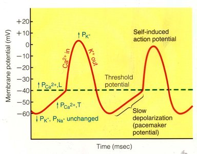
Pacemaker cells in the SA node generate spontaneous action potentials:
Slow depolarization (pacemaker potential) due to Na+ and Ca2+ influx.
Rapid depolarization when threshold is reached, followed by repolarization via K+ efflux.
Regulation of Heart Rate and Cardiac Output
Autonomic Nervous System Control
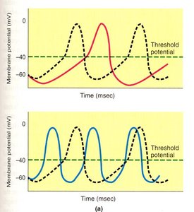
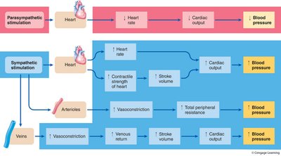
The autonomic nervous system (ANS) modulates heart rate and contractility:
Parasympathetic (Vagus Nerve): Releases ACh, slows heart rate by hyperpolarizing pacemaker cells.
Sympathetic: Releases norepinephrine, increases heart rate and contractility by depolarizing pacemaker cells.
Cardiac Output
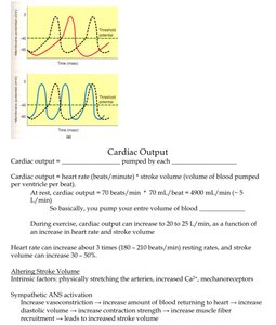
Cardiac output (CO) is the volume of blood pumped by each ventricle per minute:
Formula: $\text{CO} = \text{HR} \times \text{SV}$
Heart rate (HR): Beats per minute.
Stroke volume (SV): Volume of blood pumped per beat.
CO increases during exercise due to increased HR and SV.
Blood Pressure Regulation
Vascular Tone and Blood Flow
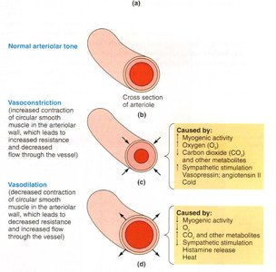
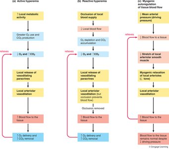
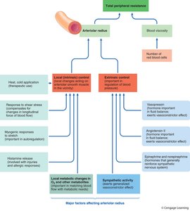
Arteriolar smooth muscle maintains vascular tone, regulating blood flow and resistance:
Vasoconstriction: Increases resistance, decreases flow.
Vasodilation: Decreases resistance, increases flow.
Regulated by local metabolic factors, sympathetic stimulation, and hormones.
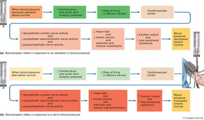
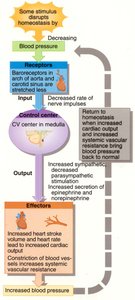
Baroreceptor Reflex
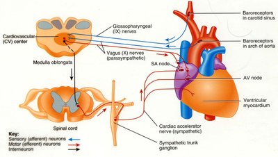
The baroreceptor reflex maintains blood pressure homeostasis:
Baroreceptors in the carotid sinus and aortic arch detect changes in blood pressure.
Signals are sent to the cardiovascular center in the medulla oblongata.
ANS adjusts heart rate, stroke volume, and vessel tone to restore normal pressure.
Mean Arterial Pressure (MAP)
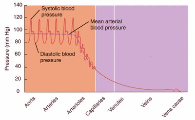
MAP is the average pressure in the arteries, driving blood flow:
Formula: $\text{MAP} = \text{CO} \times \text{TPR}$
CO: Cardiac output; TPR: Total peripheral resistance.
MAP is monitored by baroreceptors and regulated by the ANS.
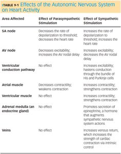
Summary Table: Effects of the Autonomic Nervous System on Heart Activity
Area Affected | Parasympathetic Stimulation | Sympathetic Stimulation |
|---|---|---|
SA node | Decreases rate of depolarization to threshold; decreases heart rate | Increases rate of depolarization to threshold; increases heart rate |
AV node | Decreases excitability; increases AV nodal delay | Increases excitability; decreases AV nodal delay |
Ventricular conduction pathway | No effect | Increases excitability; hastens conduction through His and Purkinje cells |
Atrial muscle | Weakens contractility | Increases contractility |
Ventricular muscle | No effect | Increases contractility |
Adrenal medulla | No effect | Promotes secretion of epinephrine and norepinephrine |
Veins | No effect | Increases venous return, stroke volume, and cardiac output |
Additional info: These notes expand on the original content with academic context, definitions, and examples to provide a comprehensive study guide for college-level anatomy and physiology students.
