 Back
BackStudy Guide: The Cardiovascular System – The Heart
Study Guide - Smart Notes
 Tailored notes based on your materials, expanded with key definitions, examples, and context.
The Cardiovascular System: The Heart
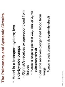
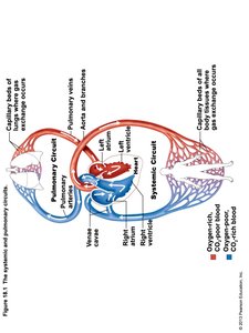
The Pulmonary and Systemic Circuits
The heart functions as a transport system, utilizing two side-by-side pumps to circulate blood throughout the body. These pumps are responsible for the pulmonary and systemic circuits, which ensure proper oxygenation and nutrient delivery.
Right side: Receives oxygen-poor blood from tissues and pumps it to the lungs via the pulmonary circuit to eliminate CO2 and absorb O2.
Left side: Receives oxygenated blood from the lungs and pumps it to body tissues via the systemic circuit.
Example: The right ventricle pumps blood to the lungs, while the left ventricle pumps blood to the rest of the body.
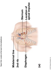
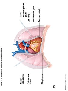
Location and Coverings of the Heart
The heart is located in the mediastinum, between the lungs, and is protected by several anatomical structures and coverings.
Location: The heart lies behind the sternum, above the diaphragm, and between the second rib and midsternal line.
Coverings: The heart is enclosed in a double-walled sac called the pericardium. The superficial fibrous pericardium anchors the heart to surrounding structures and prevents overfilling.
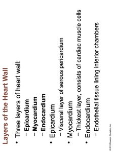
Layers of the Heart Wall
The heart wall consists of three distinct layers, each with specialized functions and cellular composition.
Epicardium: The visceral layer of the serous pericardium.
Myocardium: The thickest layer, composed of cardiac muscle cells responsible for contraction.
Endocardium: Endothelial tissue lining the interior chambers of the heart.
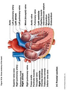
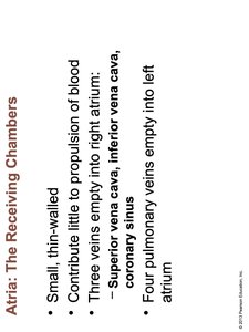
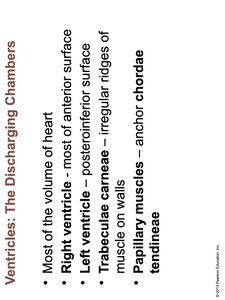
Chambers of the Heart
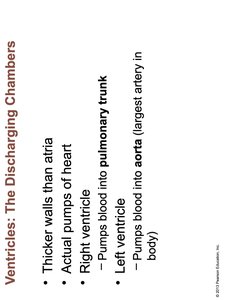
The heart contains four chambers: two atria (receiving chambers) and two ventricles (discharging chambers), each with unique structural and functional characteristics.
Atria: Small, thin-walled chambers that contribute little to propulsion of blood. The right atrium receives blood from the superior vena cava, inferior vena cava, and coronary sinus; the left atrium receives blood from four pulmonary veins.
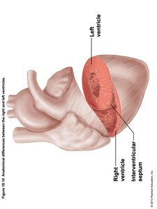
Ventricles: Larger, thicker-walled chambers that serve as the actual pumps of the heart. The right ventricle pumps blood into the pulmonary trunk; the left ventricle pumps blood into the aorta.
Structural Features: The ventricles contain trabeculae carneae (irregular ridges of muscle) and papillary muscles that anchor chordae tendineae.
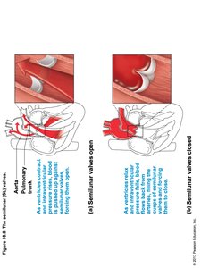
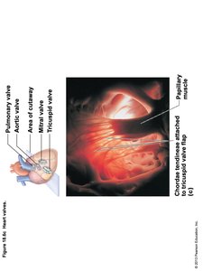
Heart Valves
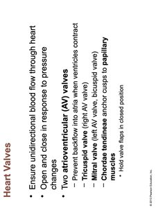
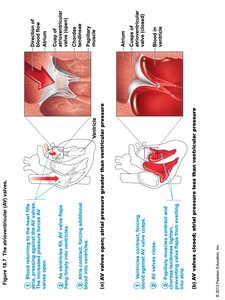
Heart valves ensure unidirectional blood flow through the heart, opening and closing in response to pressure changes. There are two main types: atrioventricular (AV) valves and semilunar (SL) valves.
Atrioventricular (AV) valves: Located between atria and ventricles; prevent backflow into atria when ventricles contract. Includes the tricuspid valve (right AV valve) and mitral valve (left AV valve).
Chordae tendineae: Anchor valve cusps to papillary muscles, holding valve flaps in closed position.
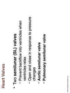
Semilunar (SL) valves: Prevent backflow into ventricles when they relax. Includes the aortic semilunar valve and pulmonary semilunar valve.
Pathway of Blood Through the Heart
Blood flows through the heart in a specific sequence, passing through valves and chambers to complete the pulmonary and systemic circuits.
Pulmonary circuit: Right atrium → tricuspid valve → right ventricle → pulmonary semilunar valve → pulmonary trunk → pulmonary arteries → lungs → pulmonary veins → left atrium.
Systemic circuit: Left atrium → mitral valve → left ventricle → aortic semilunar valve → aorta → systemic circulation.
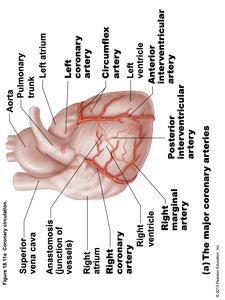
Coronary Circulation
The heart receives its own blood supply through the coronary arteries, which branch off the aorta and encircle the heart. Coronary veins return deoxygenated blood to the right atrium.
Major arteries: Right and left coronary arteries, circumflex artery, anterior and posterior interventricular arteries.
Major veins: Cardiac veins, coronary sinus.
Homeostatic Imbalances
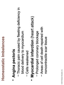
Disruptions in coronary circulation can lead to clinical conditions such as angina pectoris and myocardial infarction.
Angina pectoris: Thoracic pain caused by fleeting deficiency in blood delivery to myocardium; cells are weakened.
Myocardial infarction (heart attack): Prolonged coronary blockage; areas of cell death are repaired with noncontractile scar tissue.
Cardiac Muscle Contraction
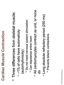
Cardiac muscle differs from skeletal muscle in several ways, including automaticity, contractile behavior, and refractory period.
Automaticity: About 1% of cells can depolarize without nervous system stimulation (autorhythmicity).
Contractile behavior: All cardiomyocytes contract as a unit, or none do.
Refractory period: Long absolute refractory period (250 ms) prevents tetanic contractions.
Pacemaker (Autorhythmic) Cells
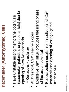
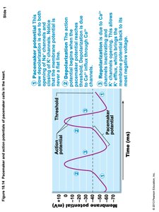
Pacemaker cells have unstable resting membrane potentials, allowing them to initiate action potentials that regulate heart rhythm.
Depolarization: Opening of slow Na+ channels causes continuous depolarization.
Threshold: Ca2+ channels open, producing the rising phase of the action potential.
Repolarization: Inactivation of Ca2+ channels and opening of voltage-gated K+ channels.
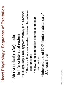
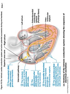
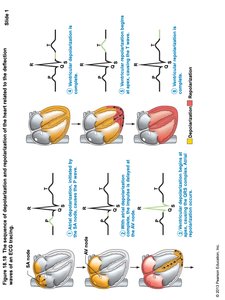
Heart Physiology: Sequence of Excitation
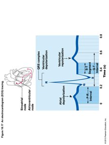
The heart's electrical activity is coordinated by a sequence of excitation, beginning with the sinoatrial (SA) node and progressing through the atrioventricular (AV) node and other conduction pathways.
SA node: Pacemaker of the heart, located in the right atrial wall; generates impulses about 75 times per minute (sinus rhythm).
AV node: Located in the inferior interatrial septum; delays impulses approximately 0.1 second, allowing atrial contraction prior to ventricular contraction.
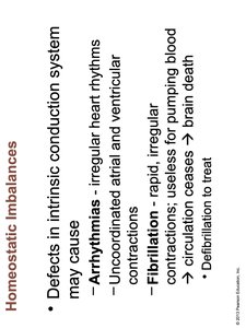
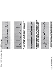
Homeostatic Imbalances: Arrhythmias and Fibrillation
Defects in the intrinsic conduction system can cause arrhythmias and fibrillation, which disrupt normal heart rhythm and function.
Arrhythmias: Irregular heart rhythms; uncoordinated atrial and ventricular contractions.
Fibrillation: Rapid, irregular contractions; useless for pumping blood, leading to circulation cessation and brain death.
Treatment: Defibrillation is used to restore normal rhythm.
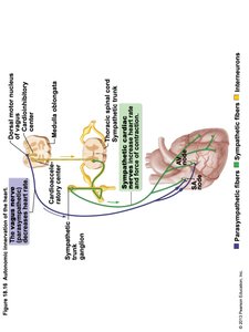
Autonomic Regulation of Heart Rate
The heart rate is regulated by the autonomic nervous system, with sympathetic and parasympathetic fibers influencing cardiac activity.
Sympathetic fibers: Increase heart rate and force of contraction.
Parasympathetic fibers: Decrease heart rate.
Electrocardiography (ECG)
An ECG records the electrical activity of the heart, providing information about depolarization and repolarization events during the cardiac cycle.
P wave: Atrial depolarization.
QRS complex: Ventricular depolarization.
T wave: Ventricular repolarization.
Mechanical Events: The Cardiac Cycle
The cardiac cycle describes the sequence of events during one complete heartbeat, including systole (contraction) and diastole (relaxation).
Systole: Contraction phase, during which blood is ejected from the chambers.
Diastole: Relaxation phase, during which chambers fill with blood.
Pressure and volume changes: Series of changes in pressure and blood volume occur throughout the cycle.
Areas for Auscultation of Heart Valves
Heart sounds can be best heard at specific intercostal spaces, corresponding to the location of the heart valves.
Aortic valve: 2nd intercostal space at right sternal margin.
Pulmonary valve: 2nd intercostal space at left sternal margin.
Mitral valve: 5th intercostal space in line with middle of clavicle.
Tricuspid valve: 5th intercostal space at right sternal margin.
Heart Chamber | Main Function | Associated Valve |
|---|---|---|
Right Atrium | Receives deoxygenated blood from body | Tricuspid Valve |
Right Ventricle | Pumps blood to lungs | Pulmonary Semilunar Valve |
Left Atrium | Receives oxygenated blood from lungs | Mitral Valve |
Left Ventricle | Pumps blood to body | Aortic Semilunar Valve |
Additional info: The notes cover the structure, function, and physiology of the heart, including its chambers, valves, conduction system, and clinical relevance. This content is directly relevant to "Ch. 18 The Cardiovascular System: The Heart" in the ANP college course.
