 Back
Study Guide: The Cardiovascular System – The Heart
Study Guide - Smart Notes
 Tailored notes based on your materials, expanded with key definitions, examples, and context.
The Cardiovascular System: The Heart
Location and Basic Structure of the Heart
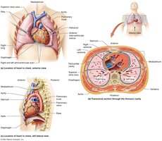
The heart is a muscular organ central to the cardiovascular system, responsible for pumping blood throughout the body. It is located in the mediastinum, a subdivision of the thoracic cavity, and sits slightly to the left within the pericardial cavity.
Shape and Size: Cone-shaped, with the apex pointing toward the left hip and the base facing the posterior rib cage. It is about the size of a fist (250–350 grams).
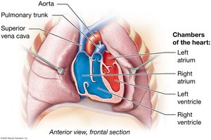
Chambers: Four hollow chambers: right and left atria (superior), right and left ventricles (inferior).
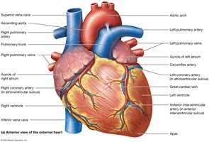
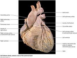
External Features: The atrioventricular sulcus marks the boundary between atria and ventricles; the interventricular sulcus separates the right and left ventricles.
Functions of the Heart: Pulmonary and Systemic Circuits
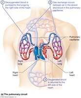
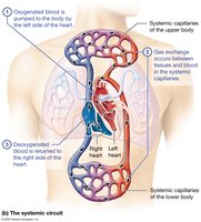
The heart operates as two pumps, each serving a distinct circuit: the pulmonary circuit (right side) and the systemic circuit (left side).
Pulmonary Circuit: Right side pumps deoxygenated blood to the lungs for gas exchange; low-pressure circuit.
Systemic Circuit: Left side pumps oxygenated blood to the body; high-pressure circuit.
Blood Flow: Veins bring blood to the heart; arteries carry blood away from the heart.
Homeostatic Functions and Endocrine Role
The heart helps maintain blood pressure and acts as an endocrine organ by producing Atrial Natriuretic Peptide (ANP), which lowers blood pressure by decreasing sodium retention in the kidneys.
Blood Pressure Regulation: Rate and force of contractions influence blood flow and pressure.
ANP: Reduces blood volume and pressure by promoting sodium and water excretion.
The Pericardium, Heart Wall, and Heart Skeleton
Pericardium and Pericardial Cavity
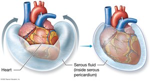
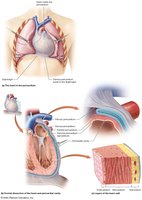
The heart is enclosed in the pericardium, a double-layered sac that protects and anchors the heart.
Fibrous Pericardium: Tough, outer layer composed of collagen bundles; anchors heart to diaphragm and great vessels.
Serous Pericardium: Thin, inner layer; consists of parietal pericardium (outer) and visceral pericardium (epicardium, inner).
Pericardial Cavity: Space between layers, filled with serous fluid to reduce friction.
Heart Wall and Skeleton
The heart wall consists of three layers, each with distinct functions.
Epicardium: Outer layer, also known as visceral pericardium.
Myocardium: Middle, thickest layer composed of cardiac muscle cells (myocytes) and fibrous skeleton.
Endocardium: Inner layer of endothelial cells and connective tissue; forms a "blood-heart barrier" regulating ion concentrations.
Fibrous Skeleton: Dense connective tissue providing structural support and electrical insulation.
The Great Vessels, Chambers, and Valves of the Heart
The Great Vessels
Major vessels associated with the heart include veins and arteries that transport blood to and from the heart.
Veins: Superior and inferior venae cavae (to right atrium), pulmonary veins (to left atrium).
Arteries: Pulmonary trunk (from right ventricle), aorta (from left ventricle).
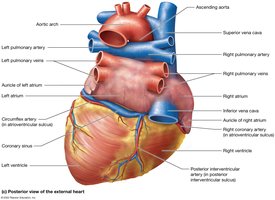
Chambers of the Heart
The heart's four chambers are divided into atria and ventricles, each with unique structural features.
Atria: Right atrium (large, thin-walled, anterior), left atrium (smaller, thicker-walled, posterior). Both have auricles for expansion.
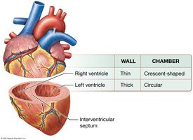
Ventricles: Right ventricle (wider, thinner walls), left ventricle (thicker walls, pumps against greater resistance).
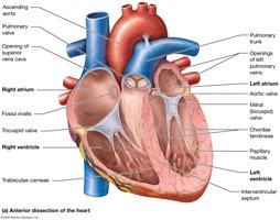
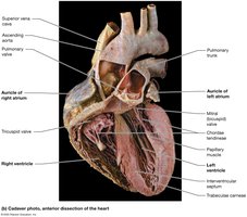
Internal Features: Pectinate muscles (right atrium), trabeculae carneae (ventricles), papillary muscles and chordae tendineae (valve support).
Septum: Interatrial and interventricular septa separate chambers.
Valves of the Heart
Valves ensure unidirectional blood flow and prevent backflow.
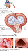
Atrioventricular (AV) Valves: Tricuspid (right), bicuspid/mitral (left); supported by chordae tendineae and papillary muscles.
Semilunar (SL) Valves: Pulmonary (right ventricle to pulmonary trunk), aortic (left ventricle to aorta); three half-moon shaped cusps.
Blood Flow Through the Heart
Overview of Blood Flow
Blood flows through the heart in a specific sequence, passing through each chamber and valve.
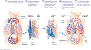
Deoxygenated blood enters the right atrium via venae cavae.
Passes through tricuspid valve to right ventricle.
Pumped through pulmonary valve to pulmonary trunk and lungs.
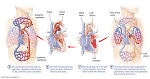
Oxygenated blood returns via pulmonary veins to left atrium.
Passes through mitral valve to left ventricle.
Pumped through aortic valve to aorta and systemic circulation.
Coronary Circulation
Coronary Arteries and Veins
The heart receives its own blood supply via the coronary circulation.
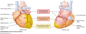
Coronary Arteries: Right and left coronary arteries branch from the ascending aorta; supply atria and ventricles.
Coronary Veins: Great, small, and middle cardiac veins drain into the coronary sinus, which empties into the right atrium.
Anastomoses: Alternate routes for blood flow; provide collateral circulation.
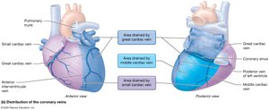
Coronary Artery Disease and Myocardial Infarction
Coronary artery disease (CAD) results from plaque buildup, leading to reduced blood flow and potential heart attack (myocardial infarction).
Symptoms: Angina pectoris, radiating chest pain, shortness of breath, nausea.
Treatments: Lifestyle changes, medications, angioplasty, stent placement, bypass grafting.
Cardiac Muscle Tissue Anatomy and Electrophysiology
Histology of Cardiac Muscle Cells
Cardiac muscle cells are striated, short, branched, and interconnected by intercalated discs containing desmosomes and gap junctions.
Functional Syncytium: Cells contract as a unit due to rapid spread of electrical activity.
Ion Channels: Voltage-gated sodium, calcium, potassium channels; HCN channels in pacemaker cells.
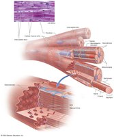
Electrophysiology: Pacemaker Cells and Conduction System
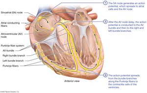
Pacemaker cells generate action potentials, setting the heart's rhythm. The conduction system includes the SA node, AV node, and Purkinje fibers.
SA Node: Fastest rate, sets sinus rhythm.
AV Node: Slower rate, conducts impulses after a delay.
Purkinje Fibers: Fast conduction to ventricles.
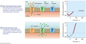
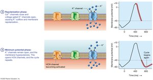
Contractile Cell Action Potentials
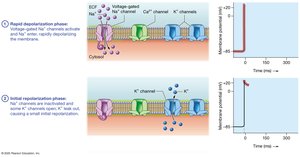
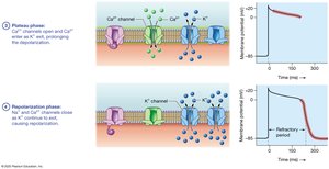
Contractile cells make up most cardiac muscle cells and have action potentials with distinct phases, including a plateau phase.
Phases: Rapid depolarization (Na+ influx), initial repolarization (K+ outflow), plateau (Ca2+ influx), repolarization (K+ outflow).
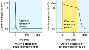
Plateau Phase: Lengthens action potential, prevents tetany, allows effective refractory period.
Electrocardiogram (ECG)
An ECG records the electrical activity of the heart, showing waves corresponding to depolarization and repolarization events.
P Wave: Atrial depolarization.
QRS Complex: Ventricular depolarization (and atrial repolarization).
T Wave: Ventricular repolarization.
Intervals: R-R (heart rate), P-R (depolarization spread), Q-T (ventricular action potentials), S-T (plateau phase).
Dysrhythmias
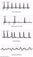
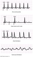
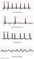
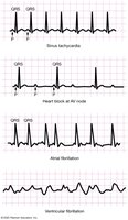
Dysrhythmias are disturbances in the heart's electrical rhythm, including bradycardia, tachycardia, heart block, and fibrillation.
Bradycardia: Heart rate < 60 bpm.
Tachycardia: Heart rate > 100 bpm.
Heart Block: Conduction delays, often at AV node.
Atrial Fibrillation: Irregular rhythm, not life-threatening.
Ventricular Fibrillation: Life-threatening, requires defibrillation.




Mechanical Physiology: The Cardiac Cycle
Cardiac Cycle and Valve Function
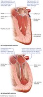
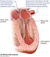
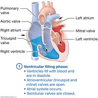
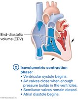
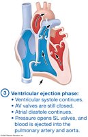
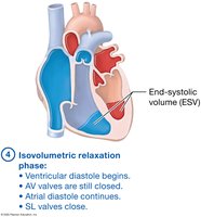
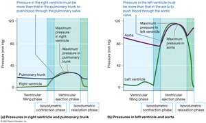
The cardiac cycle consists of alternating periods of contraction (systole) and relaxation (diastole) in the heart chambers, driven by pressure changes and valve actions.
Ventricular Filling: AV valves open, SL valves closed; ventricles fill with blood.
Isovolumetric Contraction: AV valves close, SL valves remain closed; pressure builds.
Ventricular Ejection: SL valves open, blood ejected; AV valves closed.
Isovolumetric Relaxation: SL valves close, AV valves remain closed; ventricles relax.
Heart Sounds and Murmurs
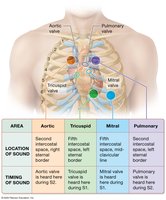
Heart sounds (S1 "lub", S2 "dub") are produced by valve closures. Murmurs indicate turbulent blood flow, often due to valve defects.
S1: AV valves close.
S2: SL valves close.
Murmurs: Audible sounds from defective valves or septal holes.

Connecting Electrical and Mechanical Events
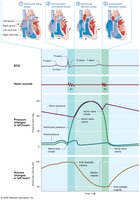
The Wiggers diagram integrates electrical, mechanical, and pressure events during the cardiac cycle, showing how ECG waves, heart sounds, pressure changes, and blood volume interact.
Cardiac Output and Regulation
Cardiac Output, Stroke Volume, and Heart Rate
Cardiac output (CO) is the volume of blood pumped by the heart per minute, determined by stroke volume (SV) and heart rate (HR).
Stroke Volume: SV = EDV - ESV (end-diastolic volume minus end-systolic volume).
Cardiac Output: CO = HR × SV
Normal Values: About 5 liters per minute for each ventricle.
Equation:
Factors Influencing Stroke Volume
Three main factors affect stroke volume: preload, contractility, and afterload.
Preload: Degree of stretch in cardiac muscle cells before contraction; determined by EDV and venous return. Frank-Starling Law: Greater stretch leads to stronger contraction.
Contractility: Ability to generate tension; influenced by inotropic agents (e.g., sympathetic stimulation).
Afterload: Resistance the ventricles must overcome to eject blood; higher afterload decreases stroke volume.
Ventricular Hypertrophy
Chronic increases in afterload can cause ventricular hypertrophy, reducing chamber size and affecting cardiac function.
Factors Influencing Heart Rate
Heart rate is primarily determined by the SA node, influenced by chronotropic agents.
Positive Chronotropic Agents: Increase heart rate (e.g., sympathetic stimulation, hormones, elevated temperature).
Negative Chronotropic Agents: Decrease heart rate (e.g., parasympathetic stimulation, decreased temperature).
Regulation of Cardiac Output
Cardiac output is regulated by nervous and endocrine systems, as well as electrolyte concentrations, temperature, age, and fitness.
Sympathetic Nervous System: Increases heart rate and contractility via norepinephrine.
Parasympathetic Nervous System: Decreases heart rate via acetylcholine.
Endocrine Regulation: Epinephrine, norepinephrine, thyroid hormone, glucagon, aldosterone, antidiuretic hormone, and ANP affect cardiac output.
Heart Failure
Heart failure is a condition where the heart cannot pump effectively, leading to pulmonary or peripheral edema.
Causes: Reduced contractility, valvular disease, cardiomyopathy, electrolyte imbalances.
Treatment: Lifestyle changes, medications, surgery; focus on increasing cardiac output and reducing congestion.
