 Back
BackStudy Guide: The Central Nervous System (CNS) – Structure, Function, and Integration
Study Guide - Smart Notes
 Tailored notes based on your materials, expanded with key definitions, examples, and context.
The Central Nervous System: Overview and Functions
Functions of the Nervous System
The nervous system is responsible for detecting, integrating, and responding to internal and external stimuli. It is divided into sensory, integrative, and motor functions:
Sensory Functions: Detection of sensations within and outside the body.
Integrative Functions: Decision-making processes, carried out exclusively by the CNS.
Motor Functions: Stimulation of muscle contractions or gland secretions.
Sensory and motor functions are performed by the peripheral nervous system (PNS), while integrative functions are exclusive to the CNS.
Basic Structure of the Brain and Spinal Cord
Brain Divisions and Functions
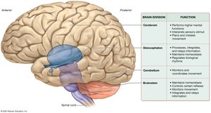
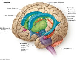
The brain is a soft, whitish-gray organ housed in the cranial cavity, composed mainly of nervous tissue. It consists of four main divisions:
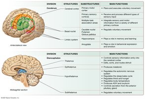
Cerebrum: Responsible for higher mental functions such as learning, memory, personality, cognition, language, and conscience. Major role in sensation and movement.
Diencephalon: Processes, integrates, and relays information; maintains homeostasis; regulates movement and biological rhythms.
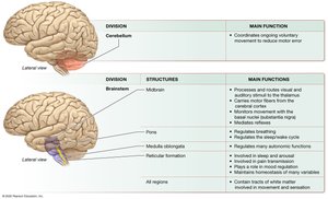
Cerebellum: Coordinates and plans movement, especially complex activities.
Brainstem: Controls basic involuntary processes, mediates reflexes, monitors movement, and relays information.
White and Gray Matter
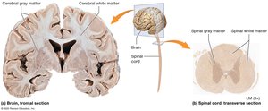
The CNS is organized into white and gray matter:
White Matter: Contains myelinated axons; forms tracts that transmit information.
Gray Matter: Composed of neuron cell bodies, dendrites, and unmyelinated axons; forms nuclei and cortex.
In the brain, gray matter is superficial and scattered in deeper regions; in the spinal cord, gray matter is internal and white matter is superficial.
Development of the CNS
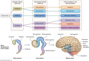
The CNS develops from the neural tube, which forms primary and secondary brain vesicles:
Primary Brain Vesicles: Forebrain, Midbrain, Hindbrain.
Secondary Brain Vesicles: Telencephalon, Diencephalon, Mesencephalon, Metencephalon, Myelencephalon.
These vesicles give rise to the mature structures of the brain.
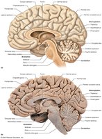
The Cerebrum
Structure and Lobes
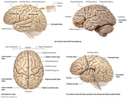
The cerebrum consists of paired hemispheres separated by the longitudinal fissure. Its surface is covered by gyri (ridges) and sulci (grooves). The main lobes are:
Frontal Lobe: Anterior, involved in motor functions and higher cognition.
Parietal Lobe: Posterior to frontal, processes sensory information.
Temporal Lobe: Lateral, involved in auditory processing and memory.
Occipital Lobe: Posterior, processes visual information.
Insula: Deep to other lobes, involved in taste and visceral sensation.
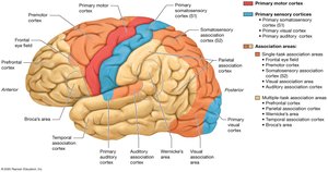
Cerebral Cortex and Functional Areas
The cerebral cortex (neocortex) is the site of conscious processes and is organized into six layers. Key functional areas include:
Primary Motor Cortex: Plans and executes movement.
Primary Sensory Cortices: Process sensory input.
Association Areas: Integrate multiple types of stimuli.
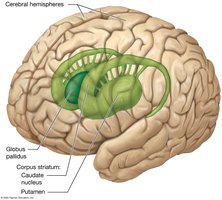
Basal Nuclei
Basal nuclei are clusters of cell bodies involved in movement regulation and behavior:
Caudate Nucleus: C-shaped, lateral to lateral ventricle.
Putamen: Posterior and inferior to caudate nucleus.
Globus Pallidus: Medial to putamen, inhibits inappropriate movements.
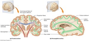
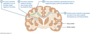
Cerebral White Matter
White matter consists of three types of fibers:
Commissural Fibers: Connect hemispheres (e.g., corpus callosum).
Projection Fibers: Connect cortex with other brain areas and spinal cord.
Association Fibers: Connect gyri within a hemisphere.
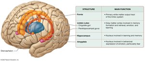
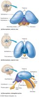
Limbic System
The limbic system is involved in memory, learning, emotion, and behavior. Major components include:
Limbic Lobe: Cingulate and parahippocampal gyri.
Hippocampus: Memory formation.
Amygdala: Emotional expression, especially fear.
The Diencephalon
Structure and Components
The diencephalon is centrally located and consists of four parts:
Thalamus: Main relay station for sensory and motor information.
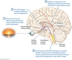
Epithalamus: Contains pineal gland, regulates sleep/wake cycle.
Hypothalamus: Regulates homeostasis, endocrine functions, and autonomic nervous system.
Subthalamus: Works with basal nuclei to control movement.
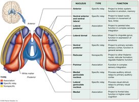
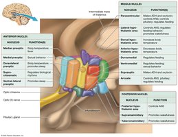
Thalamic Nuclei
The thalamus contains specific relay, association, and nonspecific nuclei, each with distinct functions in processing and integrating sensory and motor information.
Hypothalamic Nuclei
The hypothalamus connects to the pituitary gland and regulates vital functions such as thirst, hunger, sleep/wake cycle, and hormone secretion.
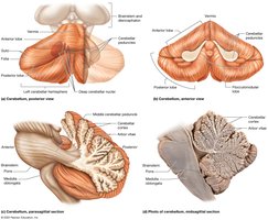
The Cerebellum
Structure and Function
The cerebellum is located posteriorly and inferiorly, divided into hemispheres and lobes. It coordinates movement and reduces motor error.
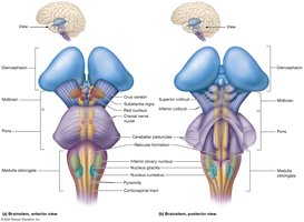
The Brainstem
Structure and Divisions
The brainstem connects the brain and spinal cord and is divided into the midbrain, pons, and medulla oblongata. It controls basic functions, mediates reflexes, and maintains alertness.
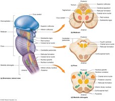
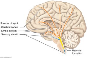
Reticular Formation
The reticular formation is a network of nuclei involved in sleep, pain regulation, mood, heart rate, ventilation, and arousal.
Integration: Major Brain Structures and Their Functions
Major brain structures work together to perform sensory, motor, integrative, and homeostatic functions.
Homeostasis and Vital Functions
Role of the Hypothalamus and Reticular Formation
The hypothalamus is the "boss" of the autonomic nervous system, adjusting output to maintain homeostasis. The reticular formation regulates heart rate, blood pressure, digestion, and urination.
Sleep and Wakefulness
Circadian Rhythms and Sleep Induction
Sleep is regulated by the suprachiasmatic nucleus (SCN) and melatonin from the epithalamus. The reticular formation and hypothalamic nuclei control sleep and arousal.
Brain Waves and Stages of Sleep
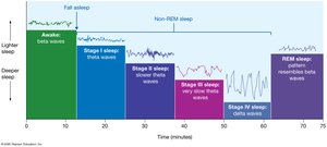
EEG measures brain waves, which change during different sleep stages:
Beta Waves: Awake, alert.
Theta Waves: Light sleep.
Delta Waves: Deep sleep.
REM Sleep: Dreaming, muscle paralysis.
Cognition, Language, Learning, and Memory
Association Areas and Lateralization
Association areas in the parietal, temporal, and prefrontal cortex are responsible for spatial awareness, recognition, personality, and moral behavior. Cognitive functions are lateralized between hemispheres.
Language Areas
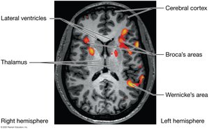
Broca's area (frontal lobe) produces language; Wernicke's area (temporal lobe) understands language. Damage results in aphasia.
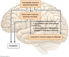
Learning and Memory
Memory is encoded and stored in neural circuits. Declarative memory involves the hippocampus and long-term potentiation (LTP); nondeclarative memory involves motor cortices, cerebellum, and basal nuclei.
Protection of the Brain and Spinal Cord
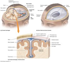
Cranial Meninges
Three protective membranes surround the brain:
Dura Mater: Outermost, tough.
Arachnoid Mater: Middle, web-like.
Pia Mater: Innermost, delicate.
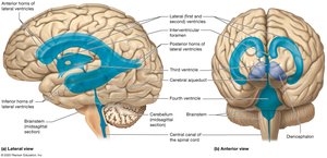
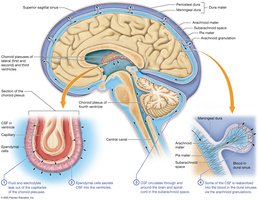
Ventricles and Cerebrospinal Fluid (CSF)
Ventricles are cavities filled with CSF, which cushions and protects the brain. CSF is produced by choroid plexuses and circulates through the ventricles and subarachnoid space.
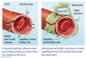
Blood Brain Barrier
The blood brain barrier consists of endothelial cells with tight junctions, limiting passage of substances from blood to brain tissue.
The Spinal Cord
Structure and Protection
The spinal cord is a relay and processing station, protected by spinal meninges and spaces (epidural, subdural, subarachnoid).
External and Internal Anatomy
The spinal cord has enlargements for limb innervation, nerve roots, and the cauda equina. Internally, it has butterfly-shaped gray matter and tracts of white matter.
Ascending and Descending Tracts
Ascending tracts carry sensory information; descending tracts carry motor commands. Key tracts include:
Posterior Columns: Touch and proprioception.
Spinothalamic Tracts: Pain and temperature.
Corticospinal Tracts: Motor control.
Sensation and Movement
Role of the CNS in Sensation
Sensory stimuli are detected by the PNS and interpreted by the CNS. General senses include touch, stretch, pain, and temperature; special senses include vision, hearing, taste, smell, and balance.
Sensory Pathways
Somatic sensory pathways include the posterior columns/medial lemniscal system and the anterolateral system. Sensory information is relayed through first-, second-, and third-order neurons to the cerebral cortex.
Somatotopy and Sensory Homunculus
The primary somatosensory cortex (S1) is organized somatotopically, with disproportionate representation for hands and face.
Role of the CNS in Voluntary Movement
Voluntary movement is planned and coordinated by motor areas of the cerebral cortex, basal nuclei, cerebellum, and spinal cord. Three types of neurons are involved: upper motor neurons, interneurons, and lower motor neurons.
Motor Homunculus
The primary motor cortex is organized somatotopically, with more area devoted to lips, tongue, and hands.
Basal Nuclei and Movement Disorders
Basal nuclei modify activity of upper motor neurons to produce voluntary movements and inhibit unintentional ones. Disorders include Parkinson's disease (hypokinetic disorder).
Cerebellum and Motor Learning
The cerebellum monitors ongoing movement, integrates sensory input, and corrects motor error. Damage results in cerebellar ataxia and intention tremor.
Integration of CNS Control of Movement
Additional info: This study guide covers the major anatomical and physiological aspects of the CNS, including its structure, function, development, protection, and integration with sensory and motor systems. It is suitable for exam preparation in college-level anatomy and physiology courses.
