 Back
BackStudy Guide: The Central Nervous System (CNS) – Structure and Function
Study Guide - Smart Notes
 Tailored notes based on your materials, expanded with key definitions, examples, and context.
The Central Nervous System
Overview and Directional Terms
The Central Nervous System (CNS) consists of the brain and spinal cord, serving as the main control center for the body. Unique directional terms are used in CNS anatomy:
Rostral: Toward the nose
Caudal: Toward the tail
The Brain
Functions of the Brain
The brain regulates vital functions and complex behaviors:
Controls: Heart rate, respiratory rate, blood pressure
Regulates: Autonomic nervous system and endocrine system
Innervates: Head via cranial nerves
Performs: Intelligence, consciousness, memory, sensory-motor integration, emotion, behavior, and socialization
Basic Parts and Organization
The brain is divided into four main regions:
Brain stem: Midbrain, pons, medulla oblongata
Cerebellum
Diencephalon: Thalamus, hypothalamus, epithalamus
Cerebrum: Two cerebral hemispheres
Ventricles of the Brain
Structure and Function
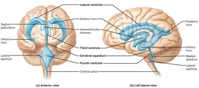
Brain ventricles are expansions of the central cavity, filled with cerebrospinal fluid (CSF) and lined with ependymal cells. They are continuous with each other and the central canal of the spinal cord.
Lateral ventricles: Located in cerebral hemispheres, horseshoe-shaped
Third ventricle: In the diencephalon, connected to lateral ventricles by interventricular foramen
Cerebral aqueduct: Connects third and fourth ventricles
Fourth ventricle: In the hindbrain, connects to central canal of spinal cord
Distribution of Gray and White Matter
Organization in the CNS
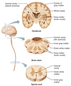
The CNS is organized with centrally located gray matter (neuronal cell bodies) and externally located white matter (axons). An additional layer of gray matter, called the cortex, is found in the cerebrum and cerebellum.
Gray matter: Contains neuronal cell bodies
White matter: Contains axons
Cortex: Outer layer of gray matter in cerebrum and cerebellum
The Brain Stem
Structure and Functions
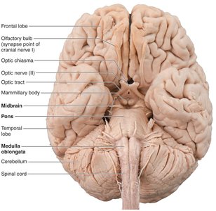
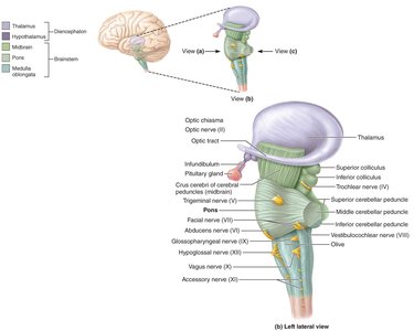
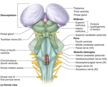
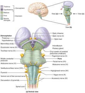
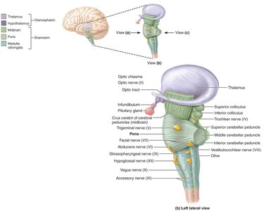
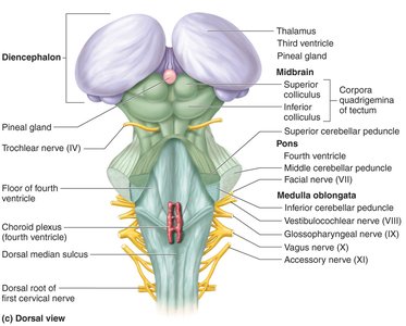
The brain stem includes the midbrain, pons, and medulla oblongata. It serves as a passageway for fiber tracts between the cerebrum and spinal cord, innervates the face and head, produces automatic behaviors necessary for survival, and integrates auditory and visual reflexes.
Medulla Oblongata
Location: Most caudal part of the brain stem, continuous with the spinal cord
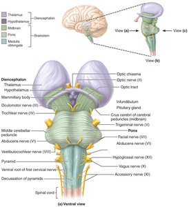
Pyramids: Major fiber tracts on ventral surface; decussation (crossing over) of motor tracts occurs here
Cranial nerves: VIII (vestibulocochlear), IX (glossopharyngeal), X (vagus), XII (hypoglossal) attach to the medulla
Reticular formation: Core contains nuclei influencing autonomic functions (cardiac, vasomotor, respiratory centers)
Pons
Bridge: Connects midbrain and medulla oblongata
Cranial nerve nuclei: V (trigeminal), VI (abducens), VII (facial)
Midbrain
Location: Between diencephalon and pons
Cerebral aqueduct: Central cavity of the midbrain
Cerebral peduncles: Contain pyramidal tracts
Corpora quadrigemina: Superior colliculi (visual reflexes), inferior colliculi (auditory reflexes)
Substantia nigra: Pigmented nuclei linked to basal nuclei, associated with Parkinson’s disease
Red nucleus: Largest nucleus of the reticular formation
The Cerebellum
Structure and Function
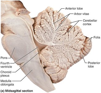
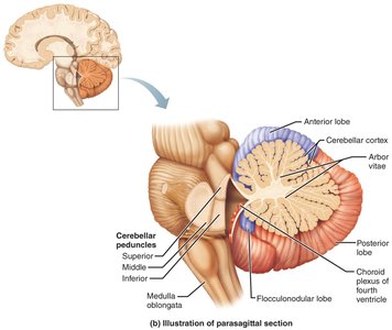
The cerebellum is located dorsal to the pons and medulla. It smooths and coordinates body movements and helps maintain equilibrium.
Hemispheres: Two, each subdivided into anterior, posterior, and flocculonodular lobes
Folia: Surface ridges separated by fissures
Regions: Cortex (gray matter), arbor vitae (internal white matter), deep cerebellar nuclei (gray matter)
Coordination of Movement
Receives information on equilibrium, current movements, and from the cerebral cortex
Compares intended movement with body position
Sends instructions to the cerebral cortex to adjust motor commands
Higher Cognitive Functions
Learning new motor skills
Participates in cognition (language, problem solving, task planning)
The Diencephalon
Structure and Components
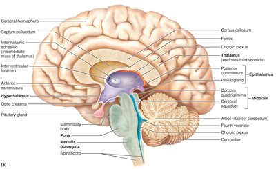
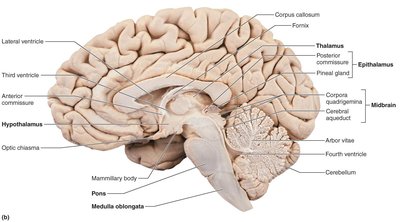
The diencephalon forms the central core of the forebrain and is composed of three paired structures: thalamus, hypothalamus, and epithalamus. It borders the third ventricle and is primarily gray matter.
Thalamus
Makes up 80% of the diencephalon
Acts as a relay station for incoming sensory messages
All communication with the cerebral cortex passes through thalamic nuclei (except olfactory information)
Organizes and amplifies or tones down signals
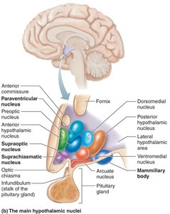
Hypothalamus
Located between optic chiasm and mammillary bodies
Pituitary gland projects inferiorly
Main visceral control center of the body
Functions: control of ANS, emotional responses, body temperature, hunger/thirst, behavior, sleep-wake cycles, endocrine system, memory formation
Epithalamus
Forms part of the roof of the third ventricle
Includes the pineal gland, which secretes melatonin and helps regulate circadian rhythm
The Cerebrum
Structure and Surface Features
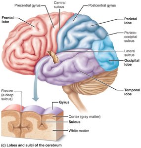
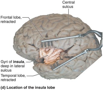
The cerebrum accounts for 83% of brain mass. Deep grooves called fissures separate major regions, while sulci (grooves) and gyri (ridges) mark the surface. Deeper sulci divide the cerebrum into lobes named for the overlying skull bones: frontal, parietal, occipital, temporal, and insula.
Cerebral Cortex
Home of conscious mind
Enables awareness, voluntary movement, communication, memory, and understanding
Composed of gray matter; folds triple its size
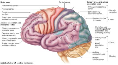
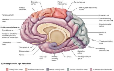
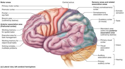
Functional regions: motor, sensory, and association areas
Functional Areas of the Cerebral Cortex
Sensory Areas
Primary sensory cortex for each major sense
Association areas process sensory information
Multimodal association areas integrate input from multiple regions
Motor Areas
Premotor cortex: controls complex movements, plans actions
Primary motor cortex: controls voluntary motor functions, located in precentral gyrus
Frontal eye field: controls voluntary eye movement
Broca’s area: manages speech production
Cerebral White Matter
Types of Tracts
Commissural fibers: Connect cerebral hemispheres (e.g., corpus callosum)
Association fibers: Connect different parts of the same hemisphere
Projection fibers: Run vertically, connecting cortex with lower regions
Deep Gray Matter of the Cerebrum
Basal Nuclei (Basal Ganglia)
Group of nuclei deep within cerebral white matter
Includes caudate nucleus, putamen, globus pallidus
Involved in motor control, start/stop/regulate voluntary movements, select appropriate muscles, estimate passage of time
Functional Brain Systems
Limbic System
Located in medial aspect of cerebral hemispheres and diencephalon
Composed of septal nuclei, cingulate gyrus, hippocampal formation, amygdaloid body
Functions: emotional processing, memory formation, pain interpretation
Reticular Formation
Runs through central core of brain stem
Reticular activating system (RAS): maintains consciousness and alertness, functions in sleep/arousal
Protection of the Brain
Meninges
Cover and protect CNS
Enclose and protect vessels supplying CNS
Contain cerebrospinal fluid between pia and arachnoid maters
Dura Mater
Strongest meninge, two layers (periosteal and meningeal)
Encloses dural sinuses
Largest sinus: superior sagittal sinus
Extends inward to subdivide cranial cavity
Arachnoid Mater
Located beneath dura mater
Arachnoid villi allow CSF to pass into dural blood sinuses
Pia Mater
Delicate connective tissue
Clings tightly to brain surface, follows cortex convolutions
Cerebrospinal Fluid (CSF)
Fills hollow cavities of brain and spinal cord
Provides liquid cushion, nourishes, removes wastes, carries chemical signals
Formed in choroid plexuses in brain ventricles
Blood-Brain Barrier
Prevents most blood-borne toxins from entering brain
Impermeable capillaries, not absolute barrier (nutrients, alcohol, nicotine, anesthetics pass through)
Spinal Cord
Structure and Function
Spinal nerves attach to it
Provides two-way conduction pathway
Major center for reflexes
Runs through vertebral canal, extends from foramen magnum to L1/L2
Key Features
Conus medullaris: inferior end
Filum terminale: connective tissue filament attaching to coccyx
Cervical/lumbar enlargements: nerves for limbs arise
Cauda equina: collection of spinal nerve roots
Spinal Cord Segments
Designated by spinal nerve issuing from it (e.g., T1)
White and Gray Matter
White matter: outer region, myelinated/nonmyelinated axons, communication between spinal cord and brain
Gray matter: shaped like "H", contains neuronal cell bodies, divided into somatic/visceral regions
Protection of the Spinal Cord
Protected by vertebrae, meninges, and CSF
Meninges: dura mater (single layer), arachnoid mater, pia mater (delicate, extends to coccyx)
Sensory and Motor Pathways in the CNS
Pathways and Tracts
Multineuron pathways connect brain and body periphery
Ascending pathways: carry information to CNS
Descending pathways: carry information away from CNS
Ascending Pathways
Conduct general somatic sensory impulses
Chains of first-, second-, and third-order neurons
Main pathways: dorsal column, spinothalamic, spinocerebellar
Descending Pathways
Deliver motor instructions from brain to spinal cord
Direct pathways (pyramidal tracts), indirect motor pathways (tectospinal, vestibulospinal, rubrospinal, reticulospinal)
Disorders of the Central Nervous System
Spinal Cord Damage
Paralysis: Loss of motor function
Parasthesia: Loss of sensation
Paraplegia: Injury between T1 and L2, paralysis of lower limbs
Quadriplegia: Injury in cervical region, paralysis of all four limbs
Brain Dysfunction
Cerebrovascular accident (stroke): Blockage/interruption of blood flow to brain region
Alzheimer’s disease: Progressive degenerative disease leading to dementia
Congenital Malformations
Anencephaly: Cerebrum and cerebellum absent
Spina bifida: Absence of vertebral lamina
Cerebral palsy: Poorly controlled voluntary muscles due to cerebral cortex damage
Postnatal Changes in the Brain
Brain structures complete development at different times
Critical periods in learning (e.g., language)
Some development continues into early 20s
Decline with age due to changes in neural circuitry and neurotransmitter release
Summary Table: Functions of Major Parts of the Brain
Brain Region | Main Function |
|---|---|
Cerebrum | Conscious thought, memory, voluntary movement |
Diencephalon | Sensory relay, homeostasis, hormone regulation |
Brain Stem | Basic life functions, cranial nerve nuclei, reflexes |
Cerebellum | Coordination, balance, motor learning |
Spinal Cord | Reflexes, sensory/motor pathway conduction |
Additional info: Table entries inferred from textbook context and lecture notes.
