 Back
BackStudy Guide: The Integumentary and Skeletal Systems
Study Guide - Smart Notes
 Tailored notes based on your materials, expanded with key definitions, examples, and context.
The Integumentary System
Overview of the Skin
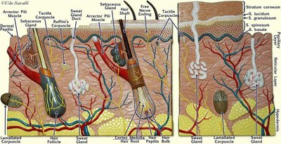
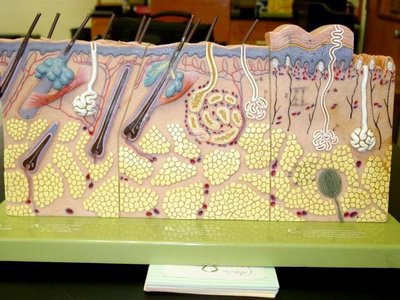
The integumentary system is composed of the skin and its derivatives, including hair, nails, and glands. The skin is the largest organ of the body and serves as a protective barrier, regulates temperature, and provides sensory information.
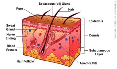
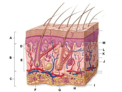
Epidermis: The outermost layer, primarily composed of keratinized stratified squamous epithelium.
Dermis: The middle layer, containing connective tissue, blood vessels, nerves, hair follicles, and glands.
Hypodermis (Subcutaneous Layer): The deepest layer, consisting mainly of adipose tissue for insulation and energy storage.
Major Structures of the Skin
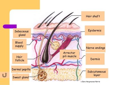
Hair Follicle: A structure in the dermis from which hair grows. Associated with sebaceous (oil) glands and arrector pili muscles.
Sebaceous Glands: Produce sebum, an oily substance that lubricates hair and skin.
Sweat Glands: Involved in thermoregulation and excretion. Two main types: eccrine (widely distributed, for cooling) and apocrine (in specific areas, associated with scent).
Arrector Pili Muscle: Smooth muscle attached to hair follicles; contraction causes "goosebumps."
Nerve Endings: Provide sensory information such as touch, pain, and temperature.
Hair Structure
Hair is a derivative of the epidermis and consists of several layers and associated structures.
Hair Shaft: The visible part of the hair above the skin surface.
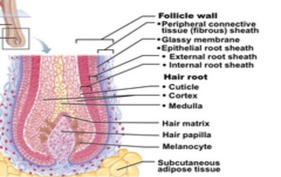
Hair Root: The portion below the surface, embedded in the follicle.
Hair Bulb: The expanded base of the hair follicle where growth occurs.
Hair Papilla: Contains blood vessels that nourish the growing hair.
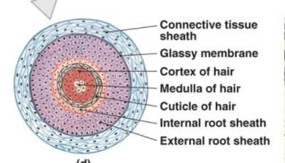
Cuticle, Cortex, Medulla: Layers of the hair shaft, with the cuticle being the outermost, cortex the middle, and medulla the innermost.
Nail Structure
Nails are protective coverings on the dorsal surface of the distal fingers and toes. They are composed of hard, keratinized cells.
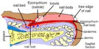
Nail Body: The visible attached part of the nail.
Nail Root: The portion embedded under the skin.
Lunula: The whitish, crescent-shaped area at the base of the nail.
Eponychium (Cuticle): The fold of skin at the base of the nail.
Hyponychium: The area under the free edge of the nail.
The Skeletal System
Overview of Bone Structure
The skeletal system provides support, protection, movement, mineral storage, and blood cell formation. Bones are classified by shape and structure, and each bone contains several anatomical features.
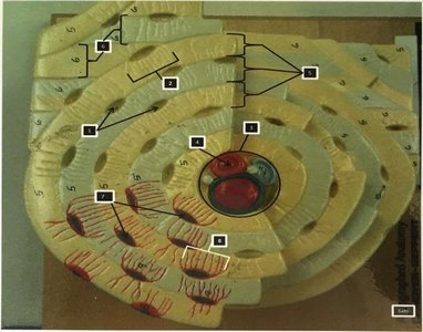
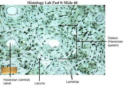
Osteon (Haversian System): The structural unit of compact bone, consisting of concentric lamellae around a central canal.
Lamellae: Rings of calcified matrix in compact bone.
Lacunae: Small spaces containing osteocytes (bone cells).
Canaliculi: Tiny channels connecting lacunae for nutrient and waste exchange.
Parts of a Long Bone
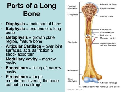
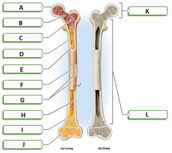
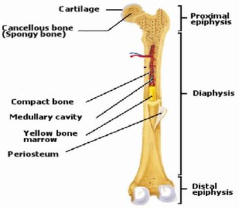
Long bones, such as the femur and humerus, have distinct regions and structures that contribute to their function.
Diaphysis: The shaft or central part of the bone.
Epiphysis: The ends of the bone, usually wider than the diaphysis.
Metaphysis: The region between the diaphysis and epiphysis; contains the growth plate in children.
Articular Cartilage: Covers joint surfaces to reduce friction and absorb shock.
Medullary Cavity: The hollow space within the diaphysis, containing bone marrow.
Endosteum: The lining of the medullary cavity.
Periosteum: The tough outer membrane covering the bone except at joint surfaces.
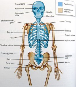
Skeletal System Overview
The human skeleton is divided into the axial and appendicular skeletons. The axial skeleton includes the skull, vertebral column, and thoracic cage, while the appendicular skeleton includes the limbs and girdles.
Axial Skeleton: Skull, vertebral column, ribs, and sternum.
Appendicular Skeleton: Pectoral girdle, pelvic girdle, and limbs.
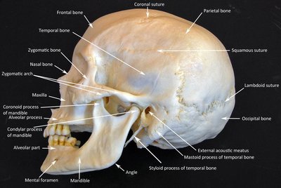
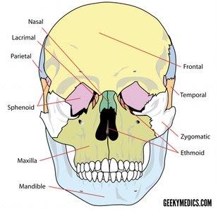
Skull Anatomy
The skull protects the brain and supports facial structures. It is composed of cranial and facial bones, with various sutures and foramina.
Cranial Bones: Frontal, parietal, temporal, occipital, sphenoid, and ethmoid bones.
Facial Bones: Maxilla, mandible, zygomatic, nasal, lacrimal, palatine, inferior nasal concha, and vomer.
Sutures: Immovable joints between skull bones (e.g., coronal, sagittal, lambdoid, squamous).
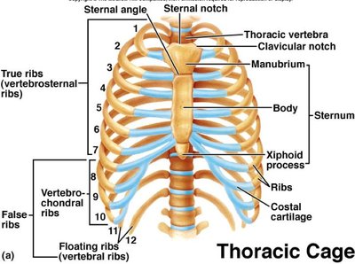
Thoracic Cage
The thoracic cage protects vital organs such as the heart and lungs and supports the shoulder girdles and upper limbs.
Sternum: Composed of the manubrium, body, and xiphoid process.
Ribs: 12 pairs; true ribs (1-7), false ribs (8-12), and floating ribs (11-12).
Costal Cartilage: Connects ribs to the sternum.
Vertebrae Structure
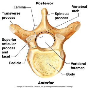
Vertebrae are the bones of the spinal column, each with characteristic features for articulation and protection of the spinal cord.
Body: The thick, disc-shaped anterior portion.
Vertebral Foramen: The opening for the spinal cord.
Spinous and Transverse Processes: Projections for muscle and ligament attachment.
Articular Processes: Form joints with adjacent vertebrae.
Pelvic Girdle (Hip)
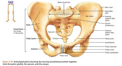
The pelvic girdle supports the weight of the upper body and protects pelvic organs. It consists of two hip bones (coxal bones), each formed by the fusion of the ilium, ischium, and pubis.
Ilium: The broad, superior part of the hip bone.
Ischium: The lower, posterior part.
Pubis: The anterior part.
Sacrum and Coccyx: Form the posterior wall of the pelvis.
Scapula and Clavicle
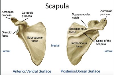
The scapula (shoulder blade) and clavicle (collarbone) form the pectoral girdle, which attaches the upper limb to the trunk.
Scapula: Features include the spine, acromion, coracoid process, and glenoid cavity.
Clavicle: S-shaped bone that articulates with the sternum and scapula.
Long Bones of the Limbs
The humerus (upper arm) and femur (thigh) are examples of long bones, each with characteristic anatomical features for articulation and muscle attachment.
Humerus: Proximal end articulates with the scapula; distal end with the radius and ulna.
Femur: The longest bone in the body; proximal end articulates with the pelvis, distal end with the tibia and patella.
Hand and Foot
The hand and foot are composed of multiple small bones that allow for a wide range of movement and dexterity.
Hand: Carpals (wrist), metacarpals (palm), phalanges (fingers).
Foot: Tarsals (ankle), metatarsals (foot), phalanges (toes).
Sutures and Sinuses of the Skull
Sutures are immovable joints between skull bones, while sinuses are air-filled cavities that reduce skull weight and resonate sound.
Major Sutures: Coronal, sagittal, lambdoid, squamous.
Sinuses: Frontal, maxillary, sphenoidal, and ethmoidal sinuses.
Synovial Joints
Synovial joints are freely movable joints characterized by a synovial cavity, articular cartilage, and a joint capsule. They allow for a wide range of movements and are found in the limbs.
Types of Synovial Joints: Hinge, ball-and-socket, pivot, saddle, plane, and condyloid.
Key Features: Articular cartilage, synovial membrane, joint cavity, ligaments.
Summary Table: Key Features of the Integumentary and Skeletal Systems
System | Main Structures | Functions |
|---|---|---|
Integumentary | Skin, hair, nails, glands | Protection, sensation, temperature regulation, excretion, vitamin D synthesis |
Skeletal | Bones, joints, cartilage, ligaments | Support, protection, movement, mineral storage, blood cell production |
