 Back
BackStudy Guide: The Muscular System (ANP College Level)
Study Guide - Smart Notes
 Tailored notes based on your materials, expanded with key definitions, examples, and context.
Muscular System Overview
Properties of Muscle Tissue
The muscular system is essential for movement, posture, and various bodily functions. Muscle tissue possesses unique properties that enable its function:
Excitability: Ability to respond to a stimulus, typically from a nerve.
Contractility: Ability to shorten forcibly when adequately stimulated.
Extensibility: Ability to extend or stretch when relaxed.
Elasticity: Ability to recoil and resume resting length after stretching.
Types of Muscle Tissue
There are three main types of muscle tissue, each with distinct characteristics, locations, and functions:
Cardiac Muscle: Found only in the heart, responsible for pumping blood, involuntary control.
Smooth Muscle: Located in walls of hollow organs (e.g., intestines, blood vessels), controls movement of substances, involuntary control.
Skeletal Muscle: Attached to bones, responsible for voluntary movements, posture, and heat generation.
Root words: Myo/Mys (muscle), Sarco (flesh)
Functions of Muscle
Produce movement
Maintain posture and body position
Stabilize joints
Generate heat
Skeletal Muscle Structure
Gross and Microscopic Anatomy
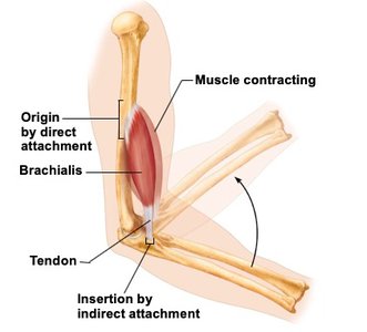
Skeletal muscles are composed of muscle fibers (cells), nerves, blood vessels, and connective tissues. Attachments to bones can be direct or indirect:
Direct attachment: Epimysium of muscle is fused to periosteum of bone or perichondrium of cartilage.
Indirect attachment: Muscle connects to bone via tendons or aponeuroses.
Muscle Fiber (Cell) Anatomy
Each muscle fiber contains specialized structures for contraction:
Sarcolemma: Plasma membrane of the muscle cell.
Sarcoplasm: Cytoplasm, rich in glycosomes and myoglobin.
Mitochondria: Numerous, providing energy.
Multinucleate: Due to fusion of embryonic cells.
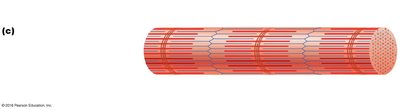
Myofibrils: Contractile elements containing myofilaments.
Sarcoplasmic reticulum: Stores calcium ions.
T tubules: Invaginations of the sarcolemma for signal transmission.
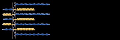
Molecular Composition of Myofilaments
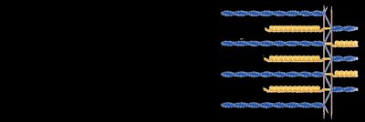
Myofibrils contain two main types of myofilaments:
Thick filaments: Composed of myosin proteins (rodlike tail, two globular heads).
Thin filaments: Composed of actin proteins (G actin polymerizes into F actin), tropomyosin, and troponin.
Elastic filament: Composed of titin, helps muscle spring back after stretching.
Dystrophin: Links thin filaments to sarcolemma proteins.
Muscle Contraction Mechanisms
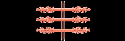
Sliding Filament Model of Contraction
Muscle contraction occurs when thin filaments slide past thick filaments, increasing overlap. The filaments themselves do not shorten; instead, the sarcomere shortens.
Myosin heads attach to actin, forming cross bridges.
Ratchet-like action pulls actin toward the center of the sarcomere.
Excitation-Contraction Coupling
This process links the electrical signal from a nerve to muscle contraction:
Stimulation by a nerve ending changes membrane potential.
Action potential is generated and propagated along the sarcolemma.
Brief rise in intracellular calcium ions triggers contraction.
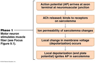
Neuromuscular Junction
The neuromuscular junction is where the motor neuron communicates with the muscle fiber:
Action potential arrives at axon terminal.
Voltage-gated Ca2+ channels open; Ca2+ enters terminal.
ACh (acetylcholine) is released and binds to receptors on sarcolemma.
Ion channels open, allowing Na+ in and K+ out.
ACh is broken down by acetylcholinesterase.

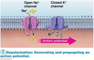
Generation of Action Potential Across the Sarcolemma
Depolarization occurs as Na+ enters and K+ exits the muscle cell, generating and propagating an action potential.
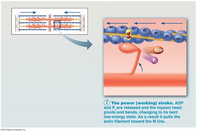
Cross Bridge Cycling
Cross bridge cycling is the sequence of events that leads to muscle contraction:
Formation: Myosin head binds to actin.
Power stroke: ADP and Pi are released, myosin head pivots, pulling actin.
Detachment: ATP binds to myosin, causing it to release actin.
Cocking: ATP is hydrolyzed, re-cocking the myosin head.


Muscle Mechanics
Principles of Muscle Contraction
The principles governing single muscle fiber contraction are similar to those for whole skeletal muscles. Muscle tension is the force exerted by muscle on an object, while load is the force exerted on the muscle by the object.
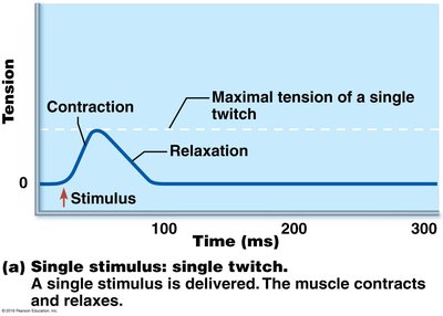
The Motor Unit and Muscle Twitch
A motor unit consists of a motor neuron and all the muscle fibers it innervates. A muscle twitch is the response to a single action potential, consisting of contraction and relaxation phases.

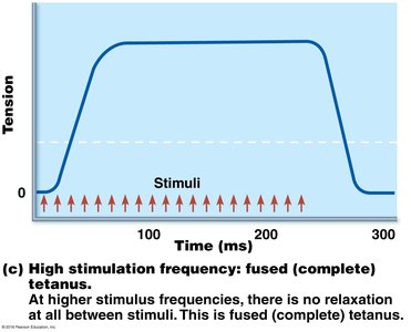
Graded Muscle Responses
Muscle contractions are graded by changing the frequency and strength of stimulation:
Frequency: Increased firing rate increases force (wave/temporal summation, tetanus).
Strength: Recruitment of more motor units increases force.
Muscle Tone
Muscle tone is a constant, slight contraction that stabilizes joints and maintains posture.
Energy for Muscle Contraction
Aerobic and Anaerobic ATP Production
ATP is required for muscle contraction, and is regenerated by three pathways:
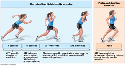
Direct phosphorylation: Creatine phosphate + ADP → ATP
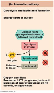
Anaerobic glycolysis: Glucose → Pyruvic acid → Lactic acid + ATP
Aerobic respiration: Glucose + O2 → CO2 + H2O + ATP

Excessive Postexercise Oxygen Consumption (EPOC)
EPOC is the extra oxygen required to restore muscles to their pre-exercise state, replenishing O2, converting lactic acid, replacing glycogen, and resynthesizing ATP and creatine phosphate.
Factors Affecting Muscle Contraction
Force of Muscle Contraction
Force depends on the number of myosin cross bridges attached to actin. Four main factors:
Number of muscle fibers recruited
Size of muscle fibers
Frequency of stimulation
Degree of muscle stretch


Velocity and Duration of Muscle Contraction
How fast and how long a muscle can contract depends on:
Muscle fiber recruitment
Load
Muscle fiber type (fast/slow, glycolytic/oxidative)
Muscle Fiber Types
Classification
Fast fibers: Rapid contraction, fatigue quickly, glycolytic pathway.
Slow fibers: Slow contraction, resistant to fatigue, oxidative pathway.
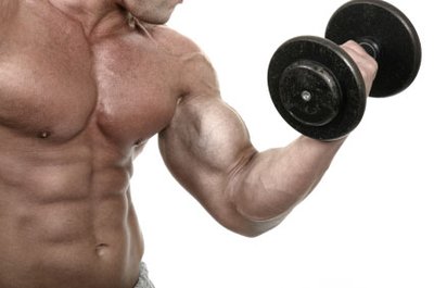
Muscle Hypertrophy
How Do Muscles Get Bigger?
Muscle hypertrophy results from high-intensity resistance exercise, typically under anaerobic conditions. Weight lifting and isometric exercise can increase muscle mass by up to 50% in a year.

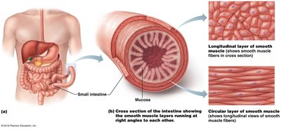
Smooth Muscle
Characteristics and Function
Smooth muscle is found in the walls of hollow organs and is responsible for involuntary movements. It is arranged in two layers, with cells oriented perpendicularly.
No striations or sarcomeres
Thick filaments shorter than thin
Filaments arranged diagonally, causing spiral contraction
Calmodulin replaces troponin for Ca2+ binding
Smooth Muscle Efficiency and Regulation
Smooth muscle contracts and relaxes 30 times longer than skeletal muscle, maintaining tension at only 1% of the energy cost. Regulation occurs via nerves, hormones, and local chemical changes.
Summary Table: Muscle Tissue Types
Type | Location | Control | Function | Structure |
|---|---|---|---|---|
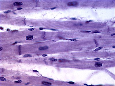
Skeletal | Attached to bones | Voluntary | Movement, posture, heat | Striated, multinucleate |
Cardiac | Heart | Involuntary | Pumping blood | Striated, branched, single nucleus |
Smooth | Walls of hollow organs | Involuntary | Movement of substances | No striations, single nucleus |
Key Equations
ATP Regeneration:
Aerobic Respiration:
Anaerobic Glycolysis:
