 Back
Study Guide: The Muscular System (BIO-168)
Study Guide - Smart Notes
 Tailored notes based on your materials, expanded with key definitions, examples, and context.
The Muscular System
Introduction to the Muscular System
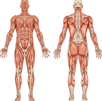
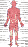
The muscular system is composed of over 600 skeletal muscles that work together to produce movement, maintain posture, and stabilize joints. Skeletal muscles attach to bones and utilize the skeleton as a system of levers, with muscle structure and arrangement determining the strength and type of movement produced.
Skeletal muscles: Voluntary muscles attached to bones.
Functions: Movement, posture, joint stabilization.
Levers: Bones act as levers, joints as fulcrums.
Interactions of Skeletal Muscles
Muscles work in groups to produce coordinated movement by pulling on bones. Each movement involves specific roles:
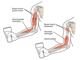
Agonist (Prime Mover): Main muscle responsible for a movement.
Antagonist: Opposes the action of the agonist.
Synergist: Assists the agonist.
Fixator: Stabilizes the origin during movement.
Example: During elbow flexion, the biceps brachii is the agonist, triceps brachii is the antagonist, brachialis is the synergist, and rotator cuff muscles act as fixators.
Agonist | Antagonist | Movement |
|---|---|---|
Biceps brachii (anterior arm) | Triceps brachii (posterior arm) | Biceps flexes forearm, triceps extends it |
Hamstring (posterior thigh) | Quadriceps femoris (anterior thigh) | Hamstrings flex leg, quadriceps extend it |
Flexor digitorum superficialis/profundus (anterior forearm) | Extensor digitorum (posterior forearm) | Flexors flex fingers/hand, extensors extend fingers/hand |
Fascicle Arrangement in Skeletal Muscles
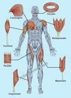
The arrangement of fascicles (bundles of muscle fibers) influences a muscle’s force and range of motion. Different patterns allow for specialized functions:
Parallel: Fibers run along the axis (e.g., sartorius).
Fusiform: Spindle-shaped, nearly parallel but taper at ends (e.g., biceps brachii).
Convergent: Fibers converge toward a single tendon (e.g., pectoralis major).
Pennate: Short fibers attach to a central tendon; can be uni-, bi-, or multipennate (e.g., deltoid).
Circular: Fibers arranged in rings (e.g., orbicularis oris).
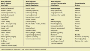
Naming Skeletal Muscles
Skeletal muscles are named based on several criteria, which help identify their location, shape, and function:
Location: e.g., brachialis (arm)
Shape: e.g., deltoid (triangle)
Size: e.g., gluteus maximus (largest)
Direction of fibers: e.g., rectus abdominus (straight)
Number of origins: e.g., biceps brachii (two heads)
Attachment points: e.g., sternocleidomastoid (sternum, clavicle, mastoid process)
Action: e.g., flexor digitorum (flexes digits)
Specific Regions | Position/Direction | Structural Characteristics | Actions |
|---|---|---|---|
Abdominis, Brachii, Capitis, Femoris | Anterior, Posterior, Medialis, Lateralis | Biceps, Triceps, Deltoid, Rectus | Flexor, Extensor, Adductor |
Axial Muscles: Head and Neck
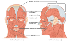
Axial muscles support and move the head, neck, and trunk. Muscles of facial expression and mastication are essential for communication and eating.
Occipitofrontalis: Raises eyebrows, wrinkles forehead.
Epicranial aponeurosis: Tendon connecting frontal and occipital bellies.
Orbicularis oris: Closes lips.
Buccinator: Compresses cheeks.
Orbicularis oculi: Closes eyelids.
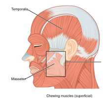
Temporalis: Elevates mandible.
Masseter: Elevates mandible.
Sternocleidomastoid: Rotates and flexes head.
Muscles Around the Eyes & Nose
These muscles control movements of the eyelids, eyebrows, and nostrils, contributing to facial expression and protection.
Orbicularis oculi: Closes eyelids.
Levator palpebrae superioris: Elevates upper eyelid.
Depressor supercilii: Pulls inner eyebrows downward.
Corrugator supercilii: Draws eyebrows toward nose.
Procerus: Pulls skin between eyebrows down.
Nasalis: Flares nostrils.
Levator labii superioris alaeque nasi: Elevates upper lip and flares nostrils.
Muscles of the Face and Mouth
Muscles in this region control movements of the lips and mouth, essential for speech and expression.
Zygomaticus major/minor: Elevate corners/upper lip.
Levator labii superioris: Elevates upper lip.
Levator anguli oris: Elevates angles of mouth.
Depressor anguli oris/labii inferioris: Depress angles/lower lip.
Risorius: Pulls angles of mouth laterally.
Mentalis: Raises central lower lip.
Platysma: Facial expression, neck movement.
The Muscles of the Eye
Six extrinsic muscles control the movement of the eyeball:
Superior rectus: Elevates eyeball.
Inferior rectus: Depresses eyeball.
Medial rectus: Moves eyeball medially.
Lateral rectus: Moves eyeball laterally.
Superior oblique: Rotates eyeball downward/laterally.
Inferior oblique: Rotates eyeball upward/laterally.
Axial Muscles: Neck
Neck muscles are divided into suprahyoid and infrahyoid groups, which support swallowing and head movement.
Suprahyoid: Digastric, stylohyoid, mylohyoid, geniohyoid.
Infrahyoid: Omohyoid, sternohyoid, thyrohyoid, sternothyroid.
Axial Muscles: Back
Back muscles stabilize and move the vertebral column and scapula.
Trapezius: Elevates, retracts, rotates scapula.
Latissimus dorsi: Extends, adducts, medially rotates arm.
Levator scapulae: Elevates scapula.
Rhomboid major/minor: Retract scapula.
Erector spinae: Extends vertebral column.
Splenius capitis: Rotates/extends neck.
Quadratus lumborum: Lateral flexion, stabilizes pelvis.
Axial Muscles: Thorax
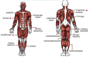
Thoracic muscles are involved in breathing and movement of the shoulder girdle.
Pectoralis major: Adducts/flexes arm.
Pectoralis minor: Pulls scapula forward/downward.
Serratus anterior: Protracts/stabilizes scapula.
Subclavius: Depresses/steadies clavicle.
External intercostals: Elevate ribs (inspiration).
Internal intercostals: Depress ribs (expiration).
Axial Muscles: Abdomen
Abdominal muscles support trunk movement, breathing, and protection of internal organs.
Diaphragm: Prime mover for inhalation.
Rectus abdominis: Flexes vertebral column, compresses abdomen.
External obliques: Rotate trunk, compress abdomen.
Internal obliques: Assist rotation/compression.
Transverse abdominis: Compresses abdominal contents.
Shoulder Muscles and Rotator Cuff
The rotator cuff stabilizes the shoulder joint and enables a wide range of arm movements.
Supraspinatus: Abducts arm.
Infraspinatus: Laterally rotates arm.
Teres minor: Laterally rotates arm.
Subscapularis: Medially rotates arm.
Teres major: Adducts, extends, medially rotates arm.
Coracobrachialis: Flexes/adducts arm.
Muscles for Forearm Movement
Muscles in the forearm are divided into anterior (flexors) and posterior (extensors) compartments.
Anterior: Biceps brachii, brachialis, brachioradialis, pronator teres.
Posterior: Triceps brachii, anconeus.
Muscles that Move the Wrist and Hand
These muscles control wrist and finger movements, essential for manipulation and grip.
Anterior (flexors): Flexor carpi radialis, palmaris longus, flexor carpi ulnaris, flexor digitorum superficialis.
Posterior (extensors): Extensor carpi radialis, extensor digitorum, extensor digiti minimi, extensor carpi ulnaris.
Muscles of the Pelvic Girdle
Muscles here move the hip and pelvis, supporting locomotion and posture.
Iliopsoas: Flexes thigh at hip.
Gluteus maximus: Extends/laterally rotates thigh.
Gluteus medius: Abducts/medially rotates thigh.
Tensor fasciae latae: Flexes/abducts thigh.
Muscles of the Thigh
Thigh muscles are grouped by compartment and function:
Anterior: Sartorius, quadriceps group (rectus femoris, vastus lateralis, vastus medialis, vastus intermedius).
Medial: Gracilis, adductor muscles, pectineus.
Posterior (Hamstrings): Biceps femoris, semitendinosus, semimembranosus.
Lower Leg Muscles
Lower leg muscles are divided into anterior, lateral, and posterior compartments, each responsible for specific movements:
Anterior: Tibialis anterior, extensor digitorum longus.
Lateral: Fibularis longus, fibularis brevis.
Posterior: Gastrocnemius, soleus, plantaris, calcaneus (Achilles) tendon.
Chapter Summary
Skeletal muscles work in coordinated groups as agonists, antagonists, synergists, and fixators.
Fascicle arrangements influence muscle strength, range of motion, and function.
Lever systems in the body determine force and movement.
Muscles are named based on location, shape, size, fiber direction, number of origins, and actions.
Axial muscles support and move the head, neck, trunk, and aid in facial expression and breathing.
Appendicular muscles move the pectoral and pelvic girdles and limbs for locomotion and manipulation.
Muscle organization reflects the integration of structure and function throughout the body.
