 Back
BackStudy Guide: The Muscular System (HKIN 162 Lab 4)
Study Guide - Smart Notes
 Tailored notes based on your materials, expanded with key definitions, examples, and context.
The Muscular System
Overview of the Muscular System
The muscular system is a complex network of tissues responsible for movement, posture, and heat generation. Skeletal muscles, which attach to bones via tendons or aponeuroses, facilitate voluntary movements by contracting and pulling on the skeleton. Most muscles work in groups to produce coordinated actions, with some acting as agonists (prime movers), antagonists, or synergists.
Agonist (Prime Mover): The main muscle responsible for a specific movement.
Antagonist: Muscle that opposes the action of the agonist.
Synergist: Muscle that assists the agonist by reducing unnecessary movement.
Example: During elbow flexion, the biceps brachii is the agonist, the triceps brachii is the antagonist, and the brachialis acts as a synergist.
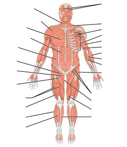
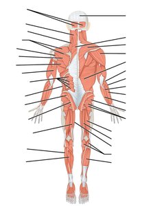
Major Muscle Groups of the Body
The body’s muscles are organized into major groups based on their location and function. Understanding these groups is essential for identifying muscle actions and their anatomical relationships.
Head and Neck Muscles: Responsible for facial expressions, mastication, and head movement (e.g., sternocleidomastoid, masseter, temporalis, orbicularis oculi, orbicularis oris).
Shoulder and Upper Arm Muscles: Move the arms and stabilize the shoulder (e.g., deltoid, biceps brachii, triceps brachii, pectoralis major, rotator cuff muscles).
Forearm and Hand Muscles: Control wrist, hand, and finger movements (e.g., flexor carpi radialis, extensor carpi ulnaris, flexor digitorum superficialis, extensor digitorum, brachioradialis).
Thoracic and Abdominal Muscles: Aid in breathing and core stability (e.g., pectoralis major, rectus abdominis, external and internal obliques, diaphragm).
Back Muscles: Support posture and allow for trunk movements (e.g., latissimus dorsi, trapezius, erector spinae, rhomboid major, levator scapulae).
Hip and Thigh Muscles: Essential for locomotion and stability (e.g., gluteus maximus, gluteus medius, quadriceps, hamstrings, adductor longus).
Leg and Foot Muscles: Facilitate walking, running, and balance (e.g., gastrocnemius, soleus, tibialis anterior, fibularis longus, extensor digitorum longus).
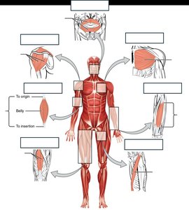
Muscle Fascicle Arrangements
Types of Muscle Fascicle Shapes
Muscle fascicles are bundles of muscle fibers arranged in specific patterns that influence a muscle’s range of motion and power. The main types are:
Parallel: Fascicles run parallel to the muscle’s long axis (e.g., sartorius, biceps brachii). High range of motion, less power.
Pennate: Fascicles attach obliquely to a central tendon. Types include:
Unipennate: Fascicles insert into one side of the tendon (e.g., extensor digitorum longus).
Bipennate: Fascicles insert into both sides of the tendon (e.g., rectus femoris).
Multipennate: Fascicles insert into multiple tendons (e.g., deltoid).
Convergent: Fascicles converge toward a single tendon (e.g., pectoralis major). Versatile movement, strong contraction.
Circular: Fascicles arranged in concentric rings (e.g., orbicularis oris, orbicularis oculi). Control openings.
Fusiform: Spindle-shaped with a thick belly (e.g., biceps brachii). Combination of range and power.
Major Muscles: Origins, Insertions, and Actions
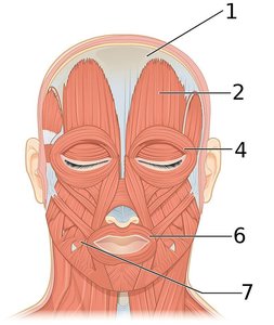
Muscles of the Face and Neck
These muscles control facial expressions, mastication, and head movement.
Muscle | Origin | Insertion | Action |
|---|---|---|---|
Frontalis | Epicranial aponeurosis | Skin superior to supraorbital margin | Draws scalp anteriorly, elevates eyebrows, wrinkles forehead |
Masseter | Maxilla and zygomatic bone | Ramus of mandible | Elevates mandible, assists in side-to-side movement, protracts mandible |
Temporalis | Temporal bone | Coronoid process and ramus of mandible | Elevates and retracts mandible, assists in side-to-side movement |
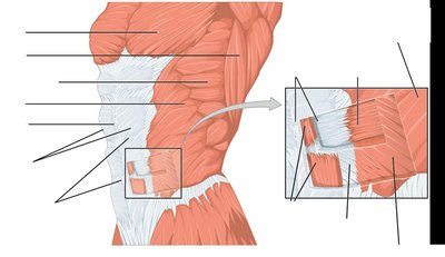
Muscles of the Abdominal Wall
The abdominal wall consists of four main muscles that flex, rotate, and compress the abdomen. The rectus abdominis is notable for its tendinous intersections, while the aponeuroses of the other muscles form the linea alba.
Muscle | Origin | Insertion | Action |
|---|---|---|---|
Rectus abdominis | Pubic crest and pubic symphysis | Cartilage of 5th–7th ribs, xiphoid process | Flexion and rotation of spine |
External oblique | 5th–12th ribs | Iliac crest, linea alba | Compresses abdomen, flexes and rotates spine |
Internal oblique | Iliac crest, inguinal ligament, thoracolumbar fascia | Cartilage of 7th–10th ribs, linea alba | Compresses abdomen, flexes and rotates spine |
Transversus abdominis | Iliac crest, inguinal ligament, lumbar fascia, cartilage of 5th–10th ribs | Xiphoid process, linea alba, pubis | Compresses abdomen |
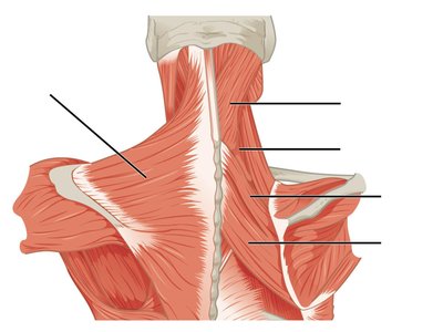
Muscles that Act on the Scapula
These muscles stabilize and move the scapula, indirectly affecting shoulder movements.
Muscle | Origin | Insertion | Action |
|---|---|---|---|
Trapezius | Occipital bone, spines of C7 and T1–T12 | Clavicle, acromion, spine of scapula | Elevates clavicle, adducts and rotates scapula, elevates/depresses scapula, extends head |
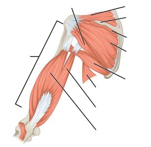
Muscles that Act on the Humerus
These muscles move and stabilize the shoulder joint, including the rotator cuff group.
Muscle | Origin | Insertion | Action |
|---|---|---|---|
Pectoralis major | Clavicle, sternum, ribs 2–6 | Greater tubercle, intertubercular sulcus of humerus | Flexes, adducts, rotates arm medially |
Latissimus dorsi | Inferior thoracic/lumbar vertebrae, sacrum, ilium, ribs 9–12 | Intertubercular sulcus of humerus | Extends, adducts, rotates arm medially |
Deltoid | Clavicle, acromion, spine of scapula | Deltoid tuberosity of humerus | Abducts, flexes, extends, rotates arm |
Rotator cuff muscles (subscapularis, supraspinatus, infraspinatus, teres minor) | Scapula | Humerus | Stabilize and move shoulder joint |
Muscles that Act on the Thigh and Leg
These muscles are responsible for movements at the hip and knee joints, including the quadriceps and hamstrings.
Muscle | Origin | Insertion | Action |
|---|---|---|---|
Quadriceps femoris (rectus femoris, vastus lateralis, vastus medialis, vastus intermedius) | Os coxae/femur | Tibial tuberosity via patellar ligament | Extend leg; rectus femoris also flexes thigh |
Hamstrings (biceps femoris, semitendinosus, semimembranosus) | Ischial tuberosity/femur | Tibia/fibula | Flex leg, extend thigh |
Gluteus maximus | Iliac crest, sacrum, coccyx | Iliotibial tract, gluteal tuberosity | Extends, rotates thigh laterally |
Gluteus medius | Ilium | Greater trochanter of femur | Abducts, rotates thigh medially |
Adductors (magnus, longus) | Pubis/ischium | Linea aspera of femur | Adduct, flex, extend, rotate thigh |
Sartorius | ASIS | Proximal medial tibia | Flexes leg, flexes and laterally rotates thigh |
Gracilis | Pubis | Medial tibia | Adducts thigh, flexes leg |
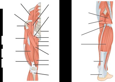
Muscles that Act on the Foot and Digits
These muscles control movements such as dorsiflexion and plantar flexion of the foot.
Muscle | Origin | Insertion | Action |
|---|---|---|---|
Gastrocnemius | Femur (lateral and medial condyles) | Calcaneus via Achilles tendon | Flexes leg, plantar flexes foot |
Soleus | Head of fibula, medial tibia | Calcaneus via Achilles tendon | Plantar flexes foot |
Muscles in Action: Functional Application
Understanding muscle actions in real movements helps reinforce anatomical knowledge. For example:
Flexing the elbow: Biceps brachii contracts, triceps brachii relaxes.
Extending the elbow: Triceps brachii contracts, biceps brachii relaxes.
Abducting the arm: Deltoid contracts.
Standing on toes: Gastrocnemius and soleus contract for plantar flexion.
Dorsiflexing the foot: Tibialis anterior contracts.
Additional info: Practicing muscle identification and movement on oneself or a volunteer reinforces the connection between anatomical structure and function.
