 Back
BackStudy Guide: The Reproductive System (Chapter 27)
Study Guide - Smart Notes
 Tailored notes based on your materials, expanded with key definitions, examples, and context.
Reproductive System Overview
Functions and General Features
The reproductive system is primarily active from puberty onward and is responsible for the formation of gametes, their union during intercourse, fertilization, and the support of fetal development and birth. Both male and female reproductive systems share homologous structures, reflecting their common embryonic origin.
Gametes: Specialized sex cells (sperm in males, oocytes in females).
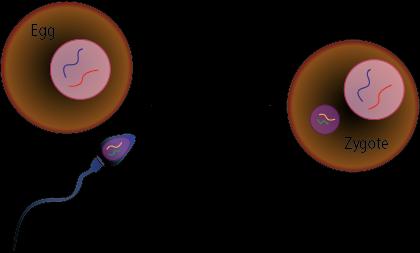
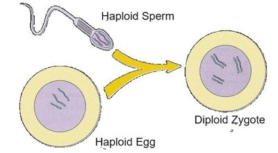
Fertilization: Fusion of sperm and egg forms a zygote.
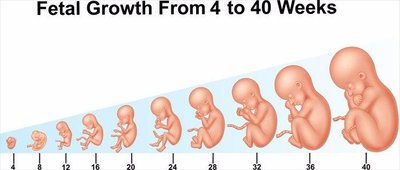
Gestation: Support and development of the fetus until birth.
Homologous Structures
Many reproductive structures in males and females are homologous, meaning they share a common origin and similar functions.
Gonads: Ovaries (female) and testes (male)
External genitalia: Penis (male) and clitoris (female)
Ducts: Vas deferens (male) and uterine tube (female)
Gonads, Gametes, and Sex Hormones
Gamete Formation
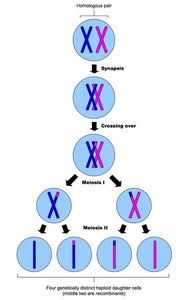
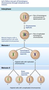
Gametes are formed in the gonads via meiosis, a specialized cell division process that reduces chromosome number by half, ensuring genetic stability across generations.
Testes: Site of sperm formation
Ovaries: Site of oocyte formation
Meiosis: Produces haploid gametes
Sex Hormones
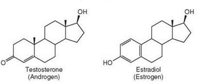
Sex hormones, mainly testosterone, estrogen, and progesterone, regulate reproductive development and secondary sexual characteristics. They are steroid-based and produced by the gonads.
Hormonal Regulation: The HPG Axis
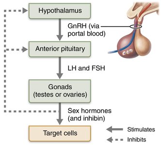
Hypothalamic-Pituitary-Gonadal (HPG) Axis
The HPG axis is a regulatory system involving the hypothalamus, pituitary gland, and gonads. It controls the secretion of sex hormones and gamete formation.
GnRH: Gonadotropin-releasing hormone from hypothalamus stimulates release of FSH and LH from anterior pituitary.
FSH: Stimulates gamete formation.
LH: Stimulates sex hormone secretion.
Inhibin: Provides negative feedback.
Meiosis and Chromosome Number
Meiosis Process
Meiosis is a two-step division process occurring only in gonads, resulting in four genetically distinct haploid cells. This ensures the zygote has the correct chromosome number after fertilization.
Humans: 46 chromosomes (23 pairs)
Homologous chromosomes: One from each parent
Sister chromatids: Copies of each chromosome
Diploid (2n): Two copies of each chromosome (all cells except gametes)
Haploid (n): One copy (gametes)

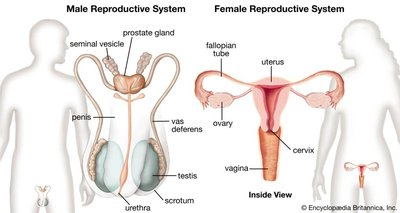
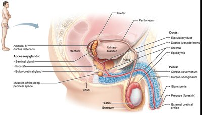
Male Reproductive System
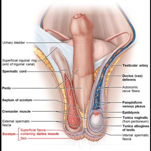
Testes and Scrotum
The testes are housed in the scrotum, which maintains a temperature lower than core body temperature for optimal sperm production. Two muscles, dartos and cremaster, regulate scrotal temperature.
Dartos muscle: Wrinkles scrotal skin to reduce heat loss.
Cremaster muscle: Elevates or lowers testes to regulate temperature.
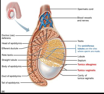
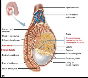
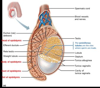
Testicular Structure
The testes are divided into lobules containing seminiferous tubules, the site of spermatogenesis. Sperm travel from seminiferous tubules to straight tubule, then to rete testis.
Tunica vaginalis: Outer covering derived from peritoneum.
Tunica albuginea: Fibrous capsule dividing testis into lobules.
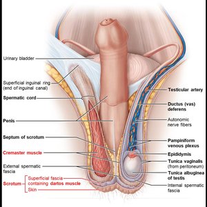
Male Duct System
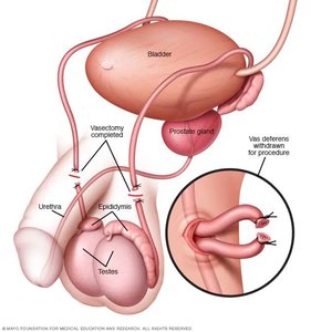
Sperm mature in the epididymis and travel through the ductus deferens, ejaculatory duct, and urethra. The ductus deferens is the site of vasectomy, a common male birth control procedure.
Epididymis: Site of sperm maturation and storage.
Ductus deferens: Transports sperm during ejaculation.
Ejaculatory duct: Formed by joining ampulla and seminal gland duct.
Urethra: Terminal duct for sperm and urine.
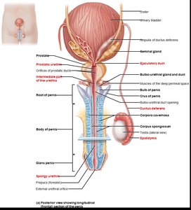
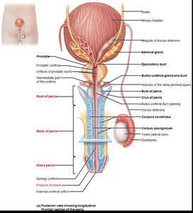
Penis and Accessory Glands
The penis is the copulatory organ, delivering sperm during intercourse. It consists of the body, glans penis, and prepuce (foreskin). Accessory glands (seminal, prostate, bulbo-urethral) produce seminal fluid.
Seminal glands: Secrete majority of seminal fluid (fructose, enzymes, prostaglandins).
Prostate gland: Secretes citrate and PSA.
Bulbo-urethral glands: Produce alkaline mucus to neutralize urine.
Semen Composition
Semen is a mixture of sperm and accessory gland secretions. It is slightly alkaline and contains nutrients, enzymes, and factors that enhance sperm motility and survival.
Fructose: Energy source for sperm.
Prostaglandins: Aid sperm movement.
Enzymes, ATP, antibiotics, clotting factors: Support sperm function.
Male Sexual Response
Erection and ejaculation are controlled by the autonomic nervous system. Erection is parasympathetic, ejaculation is sympathetic.
Erection: Nitric oxide relaxes arterioles, blood fills erectile bodies.
Ejaculation: Emission and expulsion phases, followed by resolution.
Spermatogenesis
Spermatogenesis is the process of sperm formation in the seminiferous tubules, beginning at puberty and continuing throughout life. Several cell types support this process.
Sustentocytes (Sertoli cells): Nourish and support developing sperm.
Spermatogenic cells: Give rise to sperm.
Myoid cells: Help move sperm out of tubules.
Interstitial cells (Leydig cells): Produce testosterone.
Spermatogenesis Steps
The process begins with mitosis of spermatogonia, followed by meiosis I and II, resulting in four spermatids. Spermiogenesis matures spermatids into spermatozoa.
Acrosome: Contains enzymes for egg penetration.
Body: Contains mitochondria for energy.
Tail: Flagellum for locomotion.
Role of Sustentocytes
Sustentocytes form the blood-testes barrier, provide nutrients, secrete testicular fluid, phagocytize faulty cells, and produce androgen-binding protein and inhibin.
ABP: Maintains high testosterone near spermatogenic cells.
Inhibin: Prevents further spermatogenesis.
Hormonal Regulation of Male Reproductive Function
Testosterone Activity
Testosterone secretion is regulated by the HPG axis and is essential for spermatogenesis and development of male secondary sexual characteristics.
LH: Stimulates Leydig cells to produce testosterone.
DHT and estrogen: Testosterone is converted in some organs for specific effects.
Secondary sexual characteristics: Hair growth, skin changes, voice deepening, increased bone and muscle mass.
Female Reproductive System
Ovaries and Follicle Development
The ovaries are the site of oocyte formation and sex hormone production. They are divided into cortex (follicles) and medulla (blood vessels, nerves), and anchored by ligaments.
Tunica albuginea: Surrounds ovary.
Follicles: Contain developing oocytes.
Female Duct System
The female duct system includes the uterine tubes, uterus, and vagina. The uterine tubes are the site of fertilization, and the uterus is the site of implantation and fetal development.
Uterine tubes: Infundibulum, ampulla (site of fertilization), isthmus.
Uterus: Fundus, body, cervix, endometrium (site of implantation).
Vagina: Organ of copulation, external opening covered by hymen.
External Female Genitalia
The vulva includes the mons pubis, labia majora and minora, vestibule, and clitoris. These structures are homologous to male external genitalia.
Labia majora: Homologous to testes.
Labia minora: Homologous to spongy urethra.
Clitoris: Homologous to penis.
Oogenesis and Follicle Development
Oogenesis Process
Oogenesis is the formation of oocytes, beginning during fetal development. Primary oocytes are arrested in meiosis I until puberty, and meiosis II is completed only upon fertilization.
Oogonia: Stem cells that produce primary oocytes.
Polar bodies: Non-functional cells that degenerate.
Follicles: Oocyte surrounded by pre-granulosa cells.
Follicle Development Stages
Follicle maturation takes nearly a year and involves preantral and antral phases. Dominant follicles are selected for ovulation.
Primordial follicle: Formed during fetal development.
Primary follicle: Pre-granulosa cells become cuboidal.
Secondary follicle: Granulosa cells proliferate, surrounded by theca.
Antral follicle: Fluid-filled vesicle forms, dominant follicle selected.
Ovarian and Uterine Cycles
Ovarian Cycle
The ovarian cycle consists of the follicular phase (days 1-14) and luteal phase (days 15-28). Ovulation is triggered by a surge in LH.
Follicular phase: Vesicular follicles grow, dominant follicle selected.
Luteal phase: Corpus luteum secretes progesterone and estrogen.
Uterine (Menstrual) Cycle
The uterine cycle involves cyclic changes in the endometrium, coordinated with ovarian hormone levels.
Menstrual phase (days 0-4): Shedding of functional layer.
Proliferative phase (days 5-14): Endometrium rebuilds, ovulation occurs.
Secretory phase (days 15-28): Endometrium prepares for implantation.
Birth Control and Hormonal Effects
Oral contraceptives mimic the postovulatory phase to prevent ovulation, thicken cervical mucus, and thin the uterine lining. Estrogen and progesterone promote oogenesis, follicle growth, and secondary sex characteristics.
Estrogen: Breast development, fat deposits, wider pelvis.
Progesterone: Maintains endometrial thickness.
Practice Questions
To prevent heat loss in the testes, which muscles contract?
Where are sperm produced?
What is the correct order of sperm travel?
Where does fertilization occur?
Where does implantation occur?
Which phase of the uterine cycle corresponds to the luteal phase?
Structure | Male | Female |
|---|---|---|
Gonads | Testes | Ovaries |
External Genitalia | Penis | Clitoris |
Ducts | Vas deferens | Uterine tube |
Phase | Ovarian Cycle | Uterine Cycle |
|---|---|---|
Days 1-14 | Follicular | Proliferative |
Day 14 | Ovulation | Ovulation |
Days 15-28 | Luteal | Secretory |
