 Back
Study Guide: The Reproductive System (Male and Female Anatomy & Physiology)
Study Guide - Smart Notes
 Tailored notes based on your materials, expanded with key definitions, examples, and context.
The Reproductive System
Overview of the Reproductive System
The reproductive system is responsible for producing gametes, facilitating fertilization, and supporting the development of offspring. It consists of primary sex organs (gonads) and accessory structures that aid in reproduction.
Primary sex organs: Testes (male), Ovaries (female)
Accessory sex organs: Glands, ducts, and external genitalia
The Male Reproductive System
External and Internal Anatomy
The male reproductive system includes the testes, scrotum, penis, and a series of ducts and glands. The testes are located within the scrotum, which provides a cooler environment necessary for sperm production.
Scrotum: Skin and superficial fascia surrounding the testes; maintains temperature 3°C below body temperature.
Penis: External organ for copulation and urination.
Ducts: Epididymis, ductus (vas) deferens, ejaculatory duct, urethra.
Accessory glands: Seminal vesicles, prostate, bulbo-urethral glands.
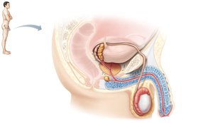
Scrotum and Testes
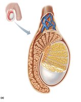
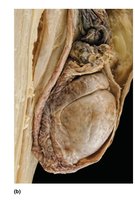
The scrotum houses the testes and is composed of skin, fascia, and muscle layers. The testes are divided into lobules containing seminiferous tubules, where sperm is produced.
Dartos muscle: Smooth muscle responsible for wrinkling scrotal skin.
Cremaster muscle: Skeletal muscle that elevates the testes.
Tunica vaginalis: Serous sac enclosing the testes.
Tunica albuginea: Fibrous capsule dividing the testis into lobules.
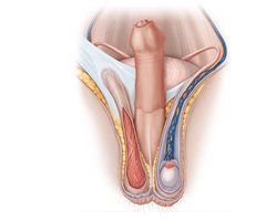
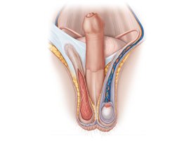
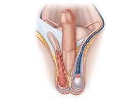
Structure of the Testis
Each testis contains 250–300 lobules, each with 1–4 seminiferous tubules. The epididymis is a comma-shaped structure on the posterior testis, essential for sperm maturation.
Seminiferous tubules: Site of spermatogenesis.
Epididymis: Stores and matures sperm.
Microscopic Anatomy of the Testis
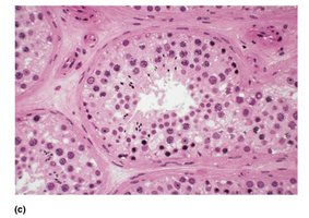
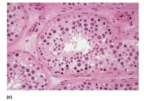
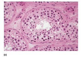
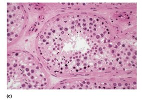
The seminiferous tubules are lined with spermatogenic cells and sustentocytes. Interstitial cells between tubules secrete testosterone.
Spermatogenic cells: Sperm-forming cells (spermatogonia, spermatocytes, spermatids).
Sustentocytes (Sertoli cells): Support and nourish developing sperm.
Myoid cells: Smooth muscle-like cells surrounding tubules.
Interstitial cells (Leydig cells): Secrete testosterone.
Spermatogenesis
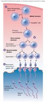
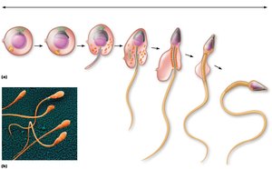
Spermatogenesis is the process of sperm formation, beginning at puberty and taking about 75 days. It involves mitosis, meiosis, and spermiogenesis.
Stage 1: Formation of spermatocytes (Type A and B cells).
Stage 2: Meiosis I and II (produces spermatids).
Stage 3: Spermiogenesis (spermatids mature into sperm).
Male Duct System
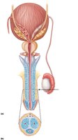
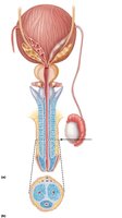
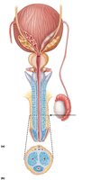
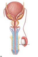
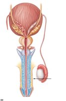
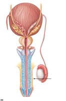
The duct system transports sperm from the testes to the outside. It includes the epididymis, ductus deferens, ejaculatory duct, and urethra.
Epididymis: Site of sperm maturation.
Ductus deferens: Stores and transports sperm; thick muscular wall.
Urethra: Carries sperm and urine to the exterior.
Accessory Glands
Accessory glands produce seminal fluid, which nourishes and protects sperm.
Seminal vesicles: Secrete fructose-rich fluid (60% of semen volume).
Prostate: Secretes fluid that enhances sperm motility (30% of semen volume).
Bulbo-urethral glands: Produce mucus to neutralize acidic urine and lubricate urethra.
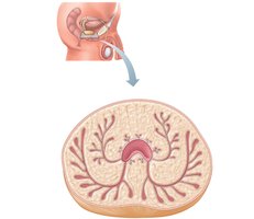
Penis: External and Internal Anatomy
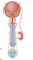
The penis consists of three erectile bodies: one corpus spongiosum and two corpora cavernosa. The glans penis is the distal expanded end, and the prepuce (foreskin) may be removed during circumcision.
Corpus spongiosum: Surrounds the spongy urethra.
Corpora cavernosa: Make up most of the penis.
Male sexual response: Erection (parasympathetic control), ejaculation (sympathetic control).
The Female Reproductive System
Overview and Anatomy
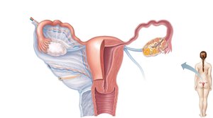
The female reproductive system produces ova, supports embryo development, and undergoes cyclical changes. It includes the ovaries, uterine tubes, uterus, vagina, and external genitalia.
Ovaries: Produce ova and hormones.
Uterine tubes: Transport oocytes; site of fertilization.
Uterus: Houses and nourishes the embryo.
Vagina: Birth canal and copulatory organ.
External genitalia: Vulva, including labia majora, labia minora, clitoris.
Oogenesis and the Menstrual Cycle
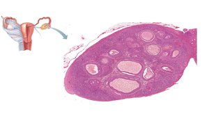
Oogenesis is the process of ovum formation, which takes many years and is completed only if fertilization occurs. The menstrual cycle is regulated by hormonal fluctuations and consists of ovarian and uterine cycles.
Oogenesis: Produces one ovum and three polar bodies; meiosis II completed only if sperm penetrates oocyte.
Ovarian cycle: Follicular phase, ovulation, luteal phase.
Uterine cycle: Menstrual phase, proliferative phase, secretory phase.
The Mammary Glands
Mammary glands are modified sweat glands responsible for milk production. Their glandular structure is undeveloped in nonpregnant women, and milk production begins at or after childbirth.
Lobes and lobules: Contain alveoli that produce milk.
Lactiferous ducts: Transport milk to the nipple.
Reproductive System Cancers
Cancers can affect both male and female reproductive organs. Early detection and treatment are crucial for prognosis.
Male: Testicular cancer (from spermatogenic cells), prostate cancer (from peripheral glands).
Female: Ovarian cancer (germinal epithelium), endometrial cancer (uterine lining), cervical cancer (HPV-related), breast cancer.
Summary Table: Male vs. Female Reproductive Structures
Male Structure | Female Structure | Function |
|---|---|---|
Testes | Ovaries | Produce gametes and hormones |
Epididymis | Uterine tubes | Sperm maturation / Oocyte transport |
Penis | Clitoris | Copulation / Erectile tissue |
Prostate, seminal vesicles | Mammary glands | Secretion of fluids |
Additional info: Hormonal regulation is essential for gametogenesis and reproductive cycles. Follicle-stimulating hormone (FSH) and luteinizing hormone (LH) play key roles in both male and female systems.
