 Back
Study Guide: The Respiratory System
Study Guide - Smart Notes
 Tailored notes based on your materials, expanded with key definitions, examples, and context.
The Respiratory System
Overview of the Respiratory System
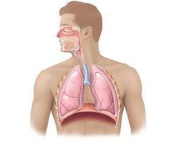
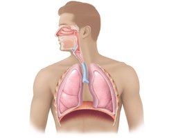
The respiratory system is responsible for the exchange of gases between the body and the external environment. It consists of several organs that work together to ensure oxygen is brought into the body and carbon dioxide is expelled. The main organs include the nose, pharynx, larynx, trachea, bronchi, and lungs (with alveoli).
Nose: Entry point for air; contains olfactory receptors and respiratory mucosa.
Pharynx: Muscular passage connecting nasal cavity to larynx; divided into nasopharynx, oropharynx, and laryngopharynx.
Larynx: Voice box; routes air and food, contains vocal cords.
Trachea: Windpipe; connects larynx to bronchi, reinforced by cartilage.
Bronchi: Branches from trachea into lungs; subdivide into smaller bronchioles.
Lungs: Main site of gas exchange; contain alveoli.
Functions of the Respiratory System
The primary function is gas exchange, which occurs in the alveoli. Passageways purify, humidify, and warm incoming air.
Gas Exchange: Oxygen enters blood, carbon dioxide exits.
Air Conditioning: Passageways filter, moisten, and warm air.
Anatomy of the Upper Respiratory Tract
The Nose
The nose is the only externally visible part of the respiratory system. Air enters through nostrils (nares) into the nasal cavity, which is divided by the nasal septum. The cavity is lined with mucosa that moistens air and traps particles.
Olfactory Receptors: Located on the superior surface for smell.
Conchae: Projections that increase surface area and air turbulence.
Palate: Separates nasal and oral cavities; anterior hard palate (bone), posterior soft palate (no bone).
Pharynx (Throat)
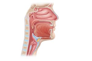
The pharynx is a muscular passageway divided into three regions: nasopharynx, oropharynx, and laryngopharynx. It serves as a common pathway for air and food.
Nasopharynx: Superior region behind nasal cavity.
Oropharynx: Middle region behind mouth.
Laryngopharynx: Inferior region attached to larynx.
Tonsils: Lymphoid tissue; pharyngeal (nasopharynx), palatine (oropharynx), lingual (base of tongue).
Larynx (Voice Box)
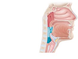
The larynx routes air and food into proper channels and plays a role in speech. It is made of eight rigid hyaline cartilages and the epiglottis (elastic cartilage).
Thyroid Cartilage: Largest cartilage; forms Adam's apple.
Epiglottis: Protects the larynx opening; closes during swallowing.
Vocal Folds: Vibrate with expelled air to produce sound; glottis is the opening.
Lower Respiratory Tract
Trachea (Windpipe)
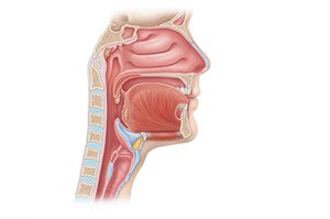
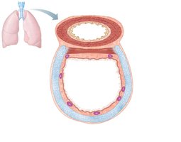
The trachea is a 4-inch tube connecting the larynx to the bronchi. Its walls are reinforced with C-shaped hyaline cartilage to keep it open. The lining contains ciliated mucosa that moves mucus and debris away from the lungs.
C-shaped Cartilage: Maintains airway patency.
Cilia: Move mucus and debris upward.
Main (Primary) Bronchi
The bronchi are formed by the division of the trachea. Each bronchus enters the lung at the hilum. The right bronchus is wider, shorter, and straighter than the left. Bronchi subdivide into smaller branches.
Lungs
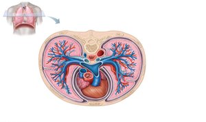
The lungs occupy most of the thoracic cavity, with the heart in the mediastinum. The apex is near the clavicle, and the base rests on the diaphragm. The left lung has two lobes, the right lung has three.
Lobes: Divided by fissures; left lung (2), right lung (3).
Mediastinum: Central area occupied by the heart.
Bronchial (Respiratory) Tree Divisions
The bronchial tree consists of branching passageways that conduct air to and from the respiratory zone. All but the smallest passageways have cartilage reinforcement.
Primary Bronchi
Secondary Bronchi
Tertiary Bronchi
Bronchioles
Terminal Bronchioles
Respiratory Zone Structures
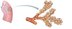
The respiratory zone is where gas exchange occurs. It includes respiratory bronchioles, alveolar ducts, alveolar sacs, and alveoli.
Alveoli: Tiny air sacs; main site of gas exchange.
Alveolar Ducts and Sacs: Pathways leading to clusters of alveoli.
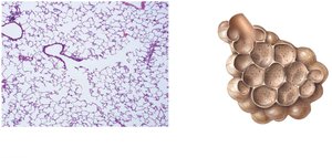
The Respiratory Membrane and Gas Exchange
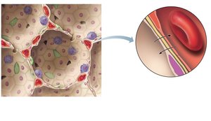
The Respiratory Membrane
Gas crosses the respiratory membrane by diffusion. Oxygen enters the blood, and carbon dioxide enters the alveoli. Alveolar macrophages provide protection by removing debris.
Structure: Composed of alveolar epithelium, fused basement membranes, and capillary endothelium.
Function: Facilitates rapid gas exchange.
Physiology of Respiration
Four Events of Respiration
Respiration involves four main events:
Pulmonary Ventilation: Moving air into and out of the lungs (breathing).
External Respiration: Gas exchange between pulmonary blood and alveoli.
Respiratory Gas Transport: Transport of oxygen and carbon dioxide via the bloodstream.
Internal Respiration: Gas exchange between blood and tissue cells in systemic capillaries.
Mechanics of Breathing (Pulmonary Ventilation)
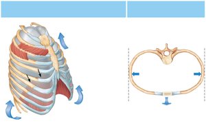
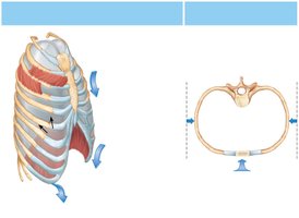
Breathing is a mechanical process dependent on volume changes in the thoracic cavity. Volume changes lead to pressure changes, causing air to flow to equalize pressure.
Inspiration (Inhalation): Active process; air flows into lungs.
Expiration (Exhalation): Passive process; air leaves lungs.
Respiratory Volumes and Capacities
Respiratory volumes and capacities describe the amount of air moved in and out of the lungs. These values are influenced by size, sex, age, and physical condition.
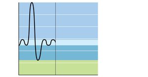
Tidal Volume (TV): Normal breath (~500 ml).
Inspiratory Reserve Volume (IRV): Extra air inhaled forcibly.
Expiratory Reserve Volume (ERV): Extra air exhaled forcibly.
Residual Volume: Air remaining after expiration.
Vital Capacity: Total exchangeable air.
Dead Space Volume: Air not reaching alveoli.
Functional Volume: Air reaching respiratory zone (~350 ml).
Respiratory capacities are measured with a spirometer.
Nonrespiratory Air Movements
Nonrespiratory movements include cough, sneeze, crying, laughing, hiccup, and yawn. These can be reflexive or voluntary and serve to clear airways or express emotion.
Respiratory Sounds
Respiratory sounds are monitored with a stethoscope. Bronchial sounds are produced by air rushing through large passageways, while vesicular sounds are softer and occur as air fills alveoli.
Gas Exchange: External and Internal Respiration
External Respiration
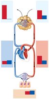
External respiration is the exchange of gases between alveoli and pulmonary capillary blood. Oxygen is loaded into the blood, and carbon dioxide is unloaded.
Diffusion: Movement toward lower concentration.
Oxygen: Moves from alveoli to blood.
Carbon Dioxide: Moves from blood to alveoli.
Gas Transport in the Blood
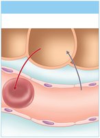
Oxygen and carbon dioxide are transported in the blood by different mechanisms.
Oxygen: Mostly attached to hemoglobin as oxyhemoglobin (HbO2); some dissolved in plasma.
Carbon Dioxide: Mostly transported as bicarbonate ion (HCO3–) in plasma; some bound to hemoglobin at different sites than oxygen.
Internal Respiration
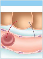
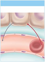
Internal respiration is the exchange of gases between blood and body cells. Oxygen is unloaded from blood to tissues, and carbon dioxide is loaded from tissues to blood.
Oxygen: Diffuses from blood into tissue cells.
Carbon Dioxide: Diffuses from tissue cells into blood.
Factors Influencing Respiratory Rate and Depth
Regulation of Breathing
Respiratory rate and depth are influenced by physical, volitional, and emotional factors.
Physical: Increased body temperature, exercise, talking, coughing.
Volitional: Conscious control.
Emotional: Fear, anger, excitement.
Summary Table: Respiratory Volumes and Capacities
Volume/Capacity | Description | Typical Value (ml) |
|---|---|---|
Tidal Volume (TV) | Normal breath | 500 |
Inspiratory Reserve Volume (IRV) | Extra air inhaled forcibly | 3,100 |
Expiratory Reserve Volume (ERV) | Extra air exhaled forcibly | 1,200 |
Residual Volume | Air remaining after expiration | 1,200 |
Vital Capacity | Total exchangeable air | 4,800 |
Total Lung Capacity | Maximum volume | 6,000 |
Key Equations
Gas Exchange (Diffusion):
Where and are partial pressures, is surface area, and is membrane thickness.
Bicarbonate Formation:
Oxygen Transport:
Carbon Dioxide Transport:
Additional info: The equations above represent the chemical reactions involved in gas transport and exchange. The rate of diffusion is influenced by partial pressure differences, surface area, and membrane thickness.
