 Back
Study Guide: The Respiratory System (BIO 169, Chapter 21)
Study Guide - Smart Notes
 Tailored notes based on your materials, expanded with key definitions, examples, and context.
The Respiratory System
Functions of the Respiratory System
The respiratory system is essential for gas exchange and maintaining homeostasis in the body. It performs several key functions:
Filters, warms, and moistens air: Prepares inhaled air for gas exchange.
Ventilation: Movement of air in and out of the lungs.
Respiration (gas exchange): Exchange of oxygen and carbon dioxide between the lungs and blood.
Speech and sound production: Facilitates vocalization.
Sense of smell: Olfactory receptors in the nasal cavity detect odors.
Maintains acid-base balance: Regulates blood pH by controlling CO2 levels.
Activation of angiotensin-II: Important for blood pressure regulation.
Classifications of the Respiratory System
The respiratory system can be classified both anatomically and physiologically:
Anatomical: Upper vs. lower respiratory tracts
Physiological: Conducting vs. respiratory zones
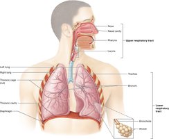
Anatomical Divisions
Upper respiratory tract: Nose, nasal cavity, pharynx, larynx
Lower respiratory tract: Trachea, bronchi, bronchioles, alveoli
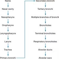
Pathway of Air
Air travels through a specific pathway in the respiratory system:
Nares → Nasal cavity → Nasopharynx → Oropharynx → Laryngopharynx → Larynx → Trachea → Primary bronchi → Secondary bronchi → Tertiary bronchi → Bronchioles → Terminal bronchioles → Respiratory bronchioles → Alveolar ducts → Alveolar sacs
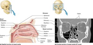
Nose and Nasal Cavity
The nose and nasal cavity filter, warm, and moisten air, and house olfactory receptors. The nasal cavity is surrounded by paranasal sinuses and contains nasal conchae and meatuses, which increase surface area and enhance resonance of voice.
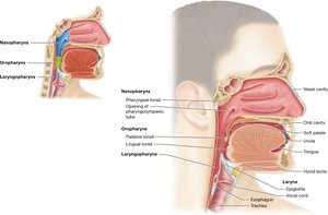
Pharynx
The pharynx is a muscular tube that connects the nasal cavity to the larynx and esophagus. It is divided into three regions: nasopharynx, oropharynx, and laryngopharynx.
Larynx
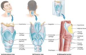
The larynx keeps food and liquid out of the respiratory tract and houses the vocal cords. It is composed of several cartilages, including the epiglottis, thyroid, and cricoid cartilages.
Trachea
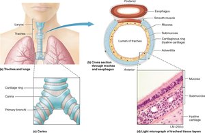
The trachea is a tube supported by C-shaped hyaline cartilage rings, which keep the airway open. Its structure includes mucosa (pseudostratified ciliated columnar epithelium with goblet cells), submucosa (loose connective tissue), and adventitia (dense irregular connective tissue).
Bronchial Tree
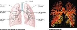
The trachea splits into right and left primary bronchi, which branch into secondary and tertiary bronchi, then into bronchioles. As the bronchi get smaller, cartilage decreases and smooth muscle increases.
Bronchioles and Alveoli
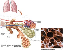
Bronchioles are small airways lacking cartilage but rich in smooth muscle. Terminal bronchioles lead to respiratory bronchioles, alveolar ducts, and alveolar sacs, which contain alveoli—the site of gas exchange.
Alveoli and Respiratory Membrane
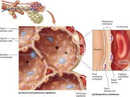
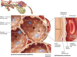
Alveoli are surrounded by elastic fibers, smooth muscle, and pulmonary capillaries. There are three cell types:
Type I alveolar cells: Simple squamous cells for gas exchange
Type II alveolar cells: Cuboidal cells that produce surfactant
Alveolar macrophages: Immune cells that remove debris
The respiratory membrane consists of Type I alveolar cells, basal lamina, and capillary endothelial cells.
The Lungs and Pleurae
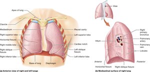
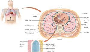
The lungs occupy the thoracic cavity except for the mediastinum. The hilum is the medial surface, and the cardiac notch is a feature of the left lung. The pleurae are double-layered serous membranes:
Parietal pleura: Outer layer
Visceral pleura: Inner layer
Pleural cavity: Space between layers containing serous fluid
Mechanics of Breathing
Steps of Respiration
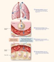
Respiration involves four steps:
Pulmonary Ventilation: Movement of air in and out of lungs
Pulmonary Gas Exchange: Exchange of gases between alveoli and blood
Gas Transport: Transport of gases through the blood
Tissue Gas Exchange: Exchange of gases between blood and cells
Pulmonary Ventilation
Pulmonary ventilation consists of two phases:
Inspiration (inhalation): Active process using diaphragm and external intercostals; increases thoracic volume
Expiration (exhalation): Passive process; relaxes muscles and decreases thoracic volume
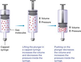
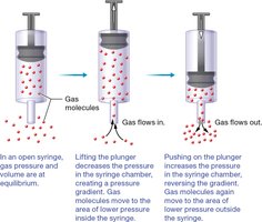
Boyle’s Law states that volume and pressure are inversely related:
Physical Factors Influencing Pulmonary Ventilation
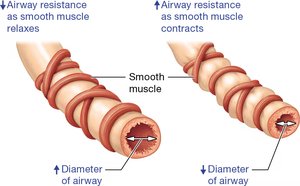
Airway resistance: Anything that impedes airflow, such as diameter, inflammation, or obstruction
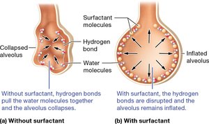
Alveolar surface tension: Alveoli are lined with water; surfactant produced by Type II cells reduces surface tension
Pulmonary compliance: Ability of lungs and chest wall to stretch
Pulmonary Volumes and Capacities
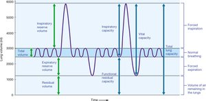
Different volumes and capacities are measured to assess lung function:
Tidal Volume (TV): 500 mL
Inspiratory Reserve Volume (IRV): 1900-3100 mL
Expiratory Reserve Volume (ERV): 700-1200 mL
Residual Volume (RV): 1100-1200 mL
Inspiratory Capacity (IC): TV + IRV
Functional Residual Capacity (FRC): ERV + RV
Vital Capacity (VC): TV + IRV + ERV
Total Lung Capacity (TLC): TV + IRV + ERV + RV
Nonrespiratory Movements
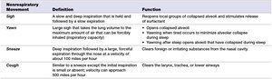
Movements such as sighs, yawns, sneezes, and coughs help keep alveoli inflated and prevent airway obstruction.
Nonrespiratory Movement | Definition | Function |
|---|---|---|
Sigh | A slow and deep inspiration that is held and followed by a slow expiration | Resets local groups of collapsed alveoli and stimulates release of surfactant |
Yawn | Long deep inspiration; opens alveoli | Opens collapsed alveoli |
Sneeze | Deep inspiration followed by a large, forceful expiration | Clears foreign or irritating substances from the nasal cavity |
Cough | Similar to sneeze but clears larynx, trachea, or lower airways | Clears lower airways |
Gas Exchange and Transport
Behavior and Movement of Gases
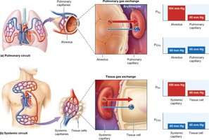
Gases move via passive diffusion from higher to lower partial pressure. Factors affecting gas exchange include partial pressures, gas solubilities, surface area, thickness of diffusion area, and perfusion.
Gas Transport
Oxygen is transported bound to hemoglobin in red blood cells. Hemoglobin saturation depends on oxygen partial pressure and affinity. The cooperative effect means that binding of one oxygen molecule increases affinity for more.
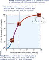
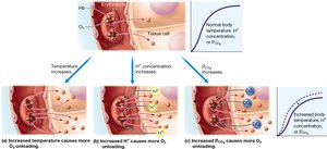
Oxygen Transport Regulation
Factors affecting hemoglobin affinity for oxygen include PO2, temperature, blood pH, PCO2, and BPG (a glycolysis product).
Carbon Dioxide Transport
CO2 is transported in three ways:
Dissolved in plasma (~7-10%)
Bound to hemoglobin (~20%)
As bicarbonate ions in blood (~70%)
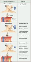
CO2 and Blood pH
Hyperventilation decreases CO2 and increases blood pH (respiratory alkalosis). Hypoventilation increases CO2 and decreases blood pH (respiratory acidosis).
Control of Ventilation
Neural Control
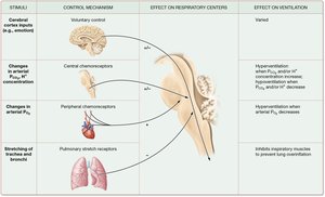
Breathing is largely autonomic, controlled by groups of neurons in the brainstem (mainly medulla oblongata). Ventral and dorsal respiratory groups regulate rhythm. Key factors include cerebral cortex/emotions, O2 and CO2 levels, H+ concentration, and stretch receptors.
Respiratory Disorders
Common Disorders
COVID-19: Viral infection affecting respiratory tract
Tuberculosis: Bacterial infection causing lung damage
COPD: Chronic obstructive pulmonary disease, includes emphysema and chronic bronchitis
Lung Cancer: Malignant growth in lung tissue
Summary Table: Steps of Respiration
Step | Description |
|---|---|
Pulmonary Ventilation | Movement of air in and out of lungs |
Pulmonary Gas Exchange | Exchange of gases between alveoli and blood |
Gas Transport | Transport of gases through the blood |
Tissue Gas Exchange | Exchange of gases between blood and cells |
Additional info: Academic context and expanded explanations were added to ensure completeness and clarity for exam preparation.
