 Back
BackStudy Guide: The Respiratory System (Chapter 22, Human Anatomy & Physiology)
Study Guide - Smart Notes
 Tailored notes based on your materials, expanded with key definitions, examples, and context.
The Respiratory System: Overview
Major Functions and Processes
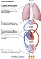
The respiratory system is essential for gas exchange, supplying oxygen to cells for cellular respiration and removing carbon dioxide. It works closely with the cardiovascular system to accomplish four key processes collectively known as respiration:
Pulmonary ventilation: Movement of air into and out of the lungs (breathing).
Pulmonary gas exchange: Exchange of gases (O2 and CO2) between lungs and blood.
Transport of respiratory gases: Movement of gases in the blood between lungs and tissues.
Tissue gas exchange: Exchange of gases between blood and body tissues.
Functional Anatomy of the Respiratory System
Major Organs and Divisions
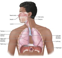
The respiratory system is divided into upper and lower regions, each with specialized structures:
Upper respiratory system: Nose, paranasal sinuses, pharynx
Lower respiratory system: Larynx, trachea, bronchi (and branches), lungs, alveoli
Respiratory muscles: Classified as part of the muscular system (e.g., diaphragm, intercostals)
The Nose and Paranasal Sinuses
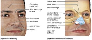
The nose is the only externally visible part of the respiratory system and serves several functions:
Provides an airway for respiration
Moistens, warms, filters, and cleans inspired air
Serves as a resonating chamber for speech
Houses olfactory receptors
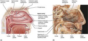
The nose is divided into the external nose and the nasal cavity. The nasal cavity is lined with mucous membranes, including olfactory mucosa (for smell) and respiratory mucosa (for air cleaning and warming).
The Pharynx
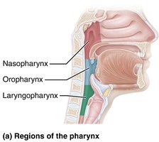
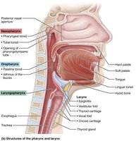
The pharynx (throat) connects the nasal cavity to the larynx and mouth to the esophagus. It is divided into three regions:
Nasopharynx: Posterior to nasal cavity, serves only as an airway
Oropharynx: Posterior to oral cavity, passageway for food and air
Laryngopharynx: Posterior to larynx, passageway for food and air
The Larynx
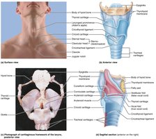
The larynx (voice box) provides an open airway, routes air and food, and houses the vocal folds for voice production. Its framework consists of nine cartilages, including the thyroid (Adam's apple), cricoid, and epiglottis.
Epiglottis: Covers the laryngeal inlet during swallowing
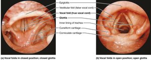
Vocal folds: Vibrate to produce sound
Vestibular folds: Help close the glottis during swallowing
The Trachea
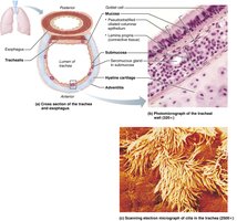
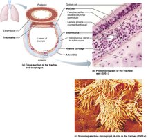
The trachea (windpipe) is a flexible tube supported by C-shaped cartilage rings. Its wall consists of three layers: mucosa, submucosa, and adventitia. The trachealis muscle contracts during coughing to expel mucus.
The Bronchi and Subdivisions
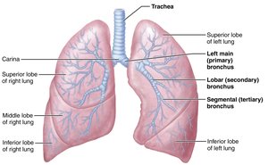
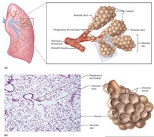
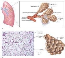
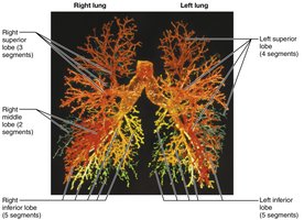
The bronchial tree undergoes about 23 generations of branching, ending in the respiratory zone structures (respiratory bronchioles, alveolar ducts, alveoli). Conducting zone structures transport air, while respiratory zone structures are sites of gas exchange.
Alveoli and the Respiratory Membrane
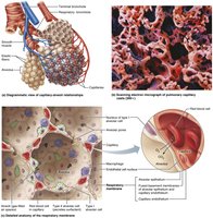
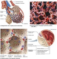
Alveoli are the primary sites of gas exchange. The respiratory membrane is extremely thin, allowing efficient diffusion. It consists of alveolar and capillary walls with a shared basement membrane. Type II alveolar cells secrete surfactant to reduce surface tension.
Gross Anatomy of the Lungs
Lung Structure and Lobes
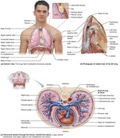
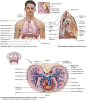
Each lung is surrounded by pleurae and connected to the mediastinum. The right lung has three lobes (superior, middle, inferior), while the left lung has two lobes (superior, inferior) and a cardiac notch to accommodate the heart.
Blood Supply and Innervation
Lungs are perfused by pulmonary and bronchial circulations. Pulmonary arteries carry deoxygenated blood to the lungs, while pulmonary veins return oxygenated blood to the heart. Bronchial arteries supply lung tissue except alveoli. Innervation is by parasympathetic (bronchoconstriction) and sympathetic (bronchodilation) fibers.
The Pleurae
Pleurae are double-layered serous membranes (parietal and visceral) that surround the lungs. Pleural fluid lubricates and creates surface tension, helping keep lungs attached to the thoracic wall.
Pulmonary Ventilation: Mechanics of Breathing
Pressure Relationships and Boyle’s Law
Pulmonary ventilation consists of inspiration and expiration. Volume changes cause pressure changes, which drive air movement. Boyle’s law describes the inverse relationship between pressure and volume:
Boyle’s Law:
Pressure Relationships in the Thoracic Cavity
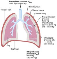
Atmospheric pressure: Pressure exerted by air surrounding the body (760 mm Hg at sea level)
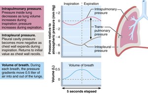
Intrapulmonary pressure: Pressure in alveoli, fluctuates during breathing
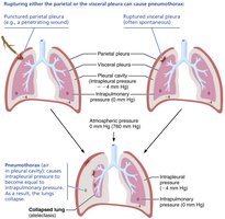
Intrapleural pressure: Pressure in pleural cavity, always negative relative to intrapulmonary pressure
Transpulmonary pressure: Difference between intrapulmonary and intrapleural pressures; keeps lungs inflated
Inspiration and Expiration
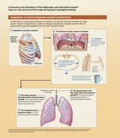
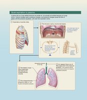
Inspiration is an active process involving contraction of the diaphragm and external intercostals, increasing thoracic volume and decreasing pressure. Expiration is usually passive, relying on relaxation and lung recoil. Forced expiration uses abdominal and internal intercostal muscles.
Physical Factors Influencing Ventilation
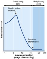
Airway resistance: Friction in airways; greatest in medium-sized bronchi
Alveolar surface tension: Surfactant reduces surface tension, preventing alveolar collapse
Lung compliance: Measure of lung distensibility; high compliance means easier ventilation
Pulmonary Volumes and Capacities
Measuring Ventilation
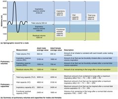
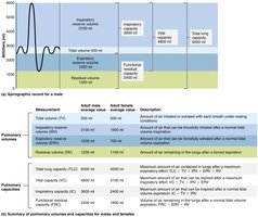
Pulmonary volumes and capacities are measured to assess respiratory status. Key volumes include:
Tidal volume (TV): Air moved in/out with each breath (~500 mL)
Inspiratory reserve volume (IRV): Air forcibly inspired beyond TV
Expiratory reserve volume (ERV): Air forcibly expired beyond TV
Residual volume (RV): Air remaining in lungs after forced expiration
Capacities are combinations of volumes:
Inspiratory capacity (IC): TV + IRV
Functional residual capacity (FRC): RV + ERV
Vital capacity (VC): TV + IRV + ERV
Total lung capacity (TLC): TV + IRV + ERV + RV
Gas Exchange: Diffusion Between Blood, Lungs, and Tissues
Basic Properties of Gases
Dalton’s Law: Total pressure of a gas mixture equals the sum of partial pressures of individual gases.
Henry’s Law: The amount of gas dissolved in a liquid is proportional to its partial pressure and solubility.
Pulmonary Gas Exchange
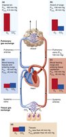
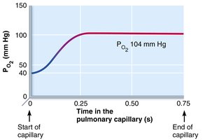
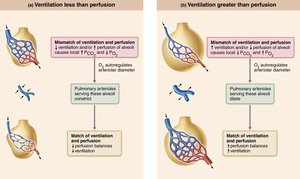
Gas exchange across the respiratory membrane is driven by partial pressure gradients and gas solubilities. Oxygen diffuses from alveoli to blood; carbon dioxide diffuses from blood to alveoli.
Tissue Gas Exchange
In systemic tissues, oxygen diffuses from blood to tissues, and carbon dioxide diffuses from tissues to blood. Venous blood returning to the heart has lower oxygen and higher carbon dioxide.
Oxygen and Carbon Dioxide Transport
Oxygen Transport
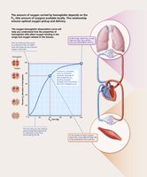
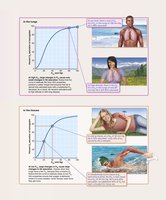
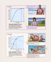
98.5% of oxygen is bound to hemoglobin (Hb) in red blood cells; 1.5% is dissolved in plasma.
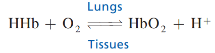
Hemoglobin can carry four O2 molecules; loading and unloading are influenced by partial pressure, temperature, pH, and BPG concentration.
Carbon Dioxide Transport
CO2 is transported in three forms: dissolved in plasma (7–10%), bound to hemoglobin (20%), and as bicarbonate ions (70%).
Formation of bicarbonate involves CO2 combining with water to form carbonic acid, which dissociates into H+ and HCO3-.
Regulation of Respiration
Neural Mechanisms
Respiratory centers in the medulla and pons control breathing rate and rhythm. The ventral respiratory group (VRG) generates the basic rhythm, while the dorsal respiratory group (DRG) integrates sensory input. Pontine centers smooth transitions between inspiration and expiration.
Chemical Factors
Arterial CO2 and pH are the most important factors affecting ventilation.
Central chemoreceptors (in brain stem) and peripheral chemoreceptors (in aortic arch and carotid arteries) detect changes.
Rising CO2 stimulates increased ventilation; low O2 becomes a major stimulus only when levels drop below 60 mm Hg.
Clinical Applications and Disorders
Common Disorders
Rhinitis: Inflammation of nasal mucosa
Laryngitis: Inflammation of vocal folds
Pleurisy: Inflammation of pleurae
Atelectasis: Lung collapse
Infant Respiratory Distress Syndrome (IRDS): Lack of surfactant in premature infants
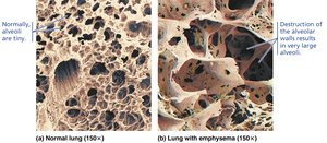
Chronic Obstructive Pulmonary Disease (COPD): Includes emphysema and chronic bronchitis
Asthma: Acute, reversible airway obstruction
Tuberculosis: Infectious disease caused by Mycobacterium tuberculosis
Lung Cancer: Leading cause of cancer deaths, often due to smoking
Sleep Apnea: Temporary cessation of breathing during sleep
Cystic Fibrosis: Genetic disorder causing thick mucus and respiratory infections
Developmental Aspects
Embryonic Development
Upper respiratory structures develop first, followed by the larynx, trachea, bronchi, and alveoli. By week 28, most premature babies can breathe on their own. Lungs continue to mature until young adulthood, and respiratory efficiency decreases with age.
Summary Table: Upper and Lower Respiratory System
Region | Main Structures | Functions |
|---|---|---|
Upper Respiratory | Nose, paranasal sinuses, pharynx | Airway, filtration, humidification, olfaction, speech resonance |
Lower Respiratory | Larynx, trachea, bronchi, lungs, alveoli | Airway, voice production, gas exchange |
Summary Table: Pulmonary Volumes and Capacities
Volume/Capacity | Definition | Average Value (Adult) |
|---|---|---|
Tidal Volume (TV) | Air moved per breath | 500 mL |
Inspiratory Reserve Volume (IRV) | Extra air inspired | 2100–3200 mL |
Expiratory Reserve Volume (ERV) | Extra air expired | 1000–1200 mL |
Residual Volume (RV) | Air remaining after forced expiration | 1200 mL |
Vital Capacity (VC) | TV + IRV + ERV | ~4800 mL |
Total Lung Capacity (TLC) | TV + IRV + ERV + RV | ~6000 mL |
Key Equations
Boyle’s Law:
Inspiratory Capacity:
Functional Residual Capacity:
Vital Capacity:
Total Lung Capacity:
Additional info:
Some clinical and developmental details were expanded for completeness.
Tables were recreated based on textbook context.
