 Back
BackStudy Guide: The Respiratory System (Chapter 22, Human Anatomy & Physiology)
Study Guide - Smart Notes
 Tailored notes based on your materials, expanded with key definitions, examples, and context.
The Respiratory System: Overview
Major Functions and Processes
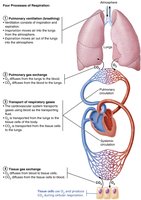
The respiratory system is essential for gas exchange, supplying oxygen to cells for cellular respiration and removing carbon dioxide, a metabolic waste. It works closely with the cardiovascular system to ensure efficient transport and exchange of gases.
Gas Exchange: The primary function is to exchange oxygen and carbon dioxide between the body and the environment.
Respiration: Involves four processes: pulmonary ventilation, pulmonary gas exchange, transport of respiratory gases, and tissue gas exchange.
Other Functions: The respiratory system also contributes to olfaction (smell) and speech.
Functional Anatomy of the Respiratory System
Major Organs and Divisions
The respiratory system is divided into upper and lower regions, each with specialized structures.
Upper Respiratory System: Includes the nose, paranasal sinuses, and pharynx.
Lower Respiratory System: Comprises the larynx, trachea, bronchi, lungs, and alveoli.
Respiratory Muscles: Classified as part of the muscular system, these muscles facilitate breathing.
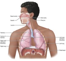
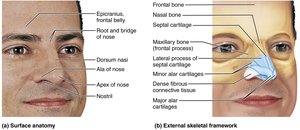
The Nose and Paranasal Sinuses
The nose is the only externally visible part of the respiratory system and serves several functions.
Airway: Provides a passage for respiration.
Moistening and Warming: Moistens and warms incoming air.
Filtering: Filters and cleans inspired air.
Speech: Acts as a resonating chamber.
Olfaction: Houses olfactory receptors.
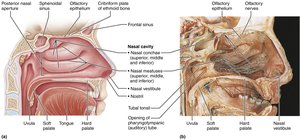
Nasal Cavity
The nasal cavity is divided by the nasal septum and lined with mucous membranes.
Olfactory Mucosa: Contains olfactory epithelium for smell.
Respiratory Mucosa: Pseudostratified ciliated columnar epithelium with goblet cells; cilia sweep mucus toward the throat.
Nasal Conchae: Increase surface area and enhance air turbulence, improving filtration and humidification.
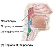
Pharynx
The pharynx connects the nasal cavity to the larynx and mouth to the esophagus. It is divided into three regions:
Nasopharynx: Airway only, lined with ciliated epithelium.
Oropharynx: Passageway for food and air, lined with stratified squamous epithelium.
Laryngopharynx: Passageway for food and air, continuous with the esophagus.
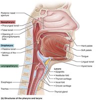
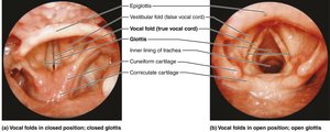
Larynx
The larynx, or voice box, is responsible for maintaining an open airway, routing air and food, and voice production.
Cartilages: Includes thyroid, cricoid, arytenoid, cuneiform, corniculate, and epiglottis.
Vocal Folds: True vocal cords vibrate to produce sound; vestibular folds (false vocal cords) help close the glottis during swallowing.
Epithelium: Stratified squamous above vocal folds; ciliated columnar below.


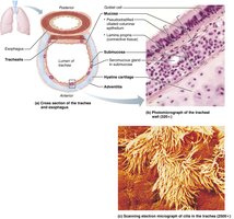
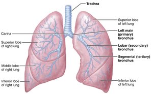
Trachea
The trachea (windpipe) is a flexible tube supported by C-shaped cartilage rings.
Layers: Mucosa (ciliated epithelium), submucosa (seromucous glands), adventitia (outer connective tissue).
Trachealis Muscle: Contracts during coughing to expel mucus.
Carina: Last cartilage marking the division into main bronchi.

Bronchi and Subdivisions
Airways branch about 23 times, forming the bronchial tree.
Main Bronchi: Right and left, entering each lung.
Lobar Bronchi: One for each lung lobe.
Segmental Bronchi: Further divisions.
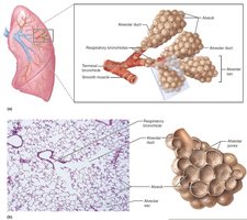
Bronchioles: Smallest branches, less than 1 mm in diameter.
Terminal Bronchioles: End of conducting zone.
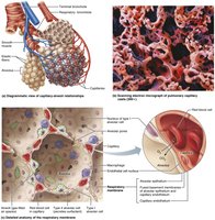
Respiratory Zone Structures
The respiratory zone includes respiratory bronchioles, alveolar ducts, and alveoli, which are the sites of gas exchange.
Alveoli: Clusters where gas exchange occurs; surrounded by capillaries and elastic fibers.
Respiratory Membrane: Thin barrier for diffusion, consisting of alveolar and capillary walls.
Surfactant: Secreted by cuboidal cells to reduce surface tension.


Lungs and Pleurae
Gross Anatomy of the Lungs
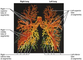
Each lung is surrounded by pleurae and divided into lobes and segments.
Right Lung: Three lobes (superior, middle, inferior).
Left Lung: Two lobes (superior, inferior), with a cardiac notch.
Bronchopulmonary Segments: Each served by its own artery, vein, and bronchus.
Lobules: Smallest subdivisions, hexagonal in shape.
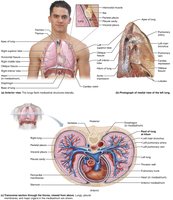

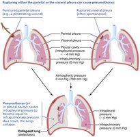
Pleurae
Pleurae are double-layered serous membranes.
Parietal Pleura: Lines thoracic wall and diaphragm.
Visceral Pleura: Covers external lung surface.
Pleural Fluid: Lubricates and creates surface tension, keeping lungs attached to thoracic wall.

Respiratory Physiology
Pulmonary Ventilation
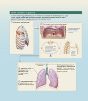
Pulmonary ventilation consists of inspiration and expiration, driven by volume and pressure changes.
Boyle’s Law: Pressure and volume of a gas are inversely related:
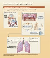
Inspiration: Active process involving diaphragm and external intercostals.
Expiration: Passive during quiet breathing; active during forced expiration.
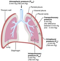
Pressure Relationships
Atmospheric Pressure: Pressure exerted by air around the body (760 mm Hg at sea level).
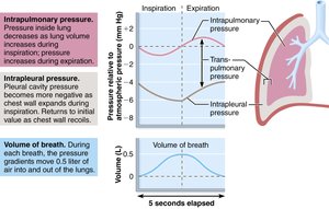
Intrapulmonary Pressure: Pressure in alveoli; fluctuates during breathing.
Intrapleural Pressure: Pressure in pleural cavity; always negative relative to intrapulmonary pressure.
Transpulmonary Pressure: Difference between intrapulmonary and intrapleural pressures; keeps lungs open.
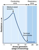
Physical Factors Influencing Ventilation
Airway Resistance: Friction in airways; greatest in medium-sized bronchi.
Alveolar Surface Tension: Surfactant reduces surface tension, preventing alveolar collapse.
Lung Compliance: Measure of lung distensibility;
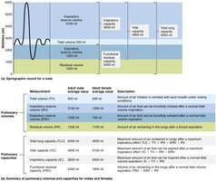
Pulmonary Volumes and Capacities
Measuring Pulmonary Function
Pulmonary volumes and capacities are assessed to evaluate respiratory health.
Tidal Volume (TV): Air moved in/out with each breath (~500 mL).
Inspiratory Reserve Volume (IRV): Air forcibly inspired beyond TV.
Expiratory Reserve Volume (ERV): Air forcibly expired beyond TV.
Residual Volume (RV): Air remaining in lungs after forced expiration.
Vital Capacity (VC):
Total Lung Capacity (TLC):

Gas Exchange
Basic Properties of Gases
Dalton’s Law: Total pressure of a gas mixture equals the sum of partial pressures of individual gases.
Henry’s Law: Gas dissolves in liquid in proportion to its partial pressure and solubility.
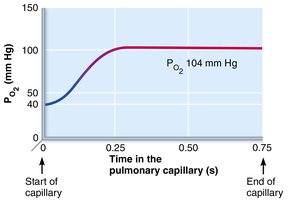
Pulmonary Gas Exchange
Gas exchange occurs across the respiratory membrane, driven by partial pressure gradients.
Oxygen: Steep gradient ensures rapid diffusion into blood.
Carbon Dioxide: Less steep gradient, but CO2 is more soluble, so diffuses efficiently.
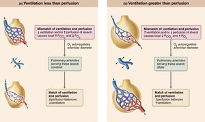
Ventilation-Perfusion Coupling: Matching of air flow and blood flow for optimal gas exchange.
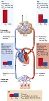

Transport of Respiratory Gases
Oxygen Transport
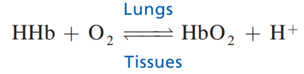
Oxygen is transported in blood primarily bound to hemoglobin.
Oxyhemoglobin: Hemoglobin bound to O2.
Deoxyhemoglobin: Hemoglobin after releasing O2.
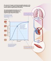
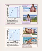
Oxygen-Hemoglobin Dissociation Curve: S-shaped curve showing relationship between partial pressure of O2 and hemoglobin saturation.

Carbon Dioxide Transport
CO2 is transported in three ways: dissolved in plasma, bound to hemoglobin, and as bicarbonate ions.
Bicarbonate Formation:
Chloride Shift: Exchange of Cl- and HCO3- across RBC membrane.
Haldane Effect: Deoxygenated hemoglobin increases CO2 carrying capacity.
Regulation of Breathing
Neural Control
Breathing is controlled by centers in the brain stem (medulla and pons).
Ventral Respiratory Group (VRG): Generates basic rhythm.
Dorsal Respiratory Group (DRG): Integrates input from peripheral receptors.
Pontine Centers: Smooth transitions between inspiration and expiration.
Chemical Regulation
Central Chemoreceptors: Respond to changes in CO2 and pH in cerebrospinal fluid.
Peripheral Chemoreceptors: Located in carotid and aortic bodies; respond to O2, CO2, and pH.
Clinical Aspects and Disorders
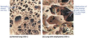
Chronic Obstructive Pulmonary Disease (COPD)
COPD includes emphysema and chronic bronchitis, characterized by irreversible airflow limitation.
Emphysema: Destruction of alveolar walls, reduced elasticity.
Chronic Bronchitis: Chronic inflammation and mucus production.
Asthma
Asthma is an acute, reversible airway obstruction due to inflammation and bronchospasm.
Tuberculosis
TB is caused by Mycobacterium tuberculosis, leading to chronic infection and lung damage.
Lung Cancer
Leading cause of cancer death; most cases are due to smoking.
Sleep Apnea
Characterized by temporary cessation of breathing during sleep, leading to daytime sleepiness and increased risk of chronic diseases.
Cystic Fibrosis
Genetic disorder causing thick mucus production, leading to airway obstruction and frequent infections.
Developmental Aspects
Embryonic Development
Upper Respiratory Structures: Develop first, followed by lower structures.
Fetal Lungs: Filled with fluid; gas exchange via placenta.
At Birth: Lungs inflate and begin functioning; respiratory rate highest in newborns.
Summary Table: Pulmonary Volumes and Capacities
Volume/Capacity | Definition | Average Value (Male) | Average Value (Female) |
|---|---|---|---|
Tidal Volume (TV) | Amount of air inhaled/exhaled with each breath | 500 mL | 500 mL |
Inspiratory Reserve Volume (IRV) | Amount of air forcibly inspired beyond TV | 3100 mL | 1900 mL |
Expiratory Reserve Volume (ERV) | Amount of air forcibly expired beyond TV | 1200 mL | 700 mL |
Residual Volume (RV) | Amount of air remaining in lungs after forced expiration | 1200 mL | 1100 mL |
Vital Capacity (VC) | TV + IRV + ERV | 4800 mL | 3100 mL |
Total Lung Capacity (TLC) | TV + IRV + ERV + RV | 6000 mL | 4200 mL |
Key Equations
Boyle’s Law:
Lung Compliance:
Oxygen-Hemoglobin Binding:
Bicarbonate Formation:
Additional info:
Some values and details inferred from standard physiology textbooks for completeness.
Tables and equations formatted for clarity and exam preparation.
