 Back
BackStudy Guide: The Skeletal System (Chapter 5) – Structure, Classification, and Function
Study Guide - Smart Notes
 Tailored notes based on your materials, expanded with key definitions, examples, and context.
The Skeletal System
Overview and Functions
The skeletal system is a complex framework of bones, joints, cartilages, and ligaments that provides structural support, protection, movement, storage, and blood cell formation. It is divided into two main subdivisions: the axial skeleton (longitudinal axis) and the appendicular skeleton (limbs and girdles).
Support: Bones form the body’s structural framework.
Protection: Bones protect vital organs (e.g., skull protects the brain).
Movement: Muscles attach to bones, enabling movement.
Storage: Bones store minerals (calcium, phosphorus) and fats.
Blood cell formation: Hematopoiesis occurs in bone marrow.
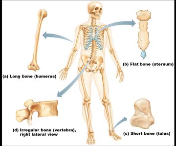
Classification of Bones
Types of Bones
Bones are classified by their shape and structure. The adult skeleton contains 206 bones, which are categorized as follows:
Long bones: Longer than they are wide; mostly compact bone (e.g., humerus).
Short bones: Cube-shaped; mostly spongy bone (e.g., talus).
Flat bones: Thin, flattened, and usually curved; two thin layers of compact bone with spongy bone in between (e.g., sternum).
Irregular bones: Complex shapes that do not fit other categories (e.g., vertebra).
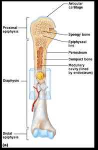
Structure of Bone
Gross Anatomy of a Long Bone
Long bones have a distinct structure, including the diaphysis (shaft), epiphyses (ends), and various membranes and cavities.
Diaphysis: Central shaft, composed of compact bone, covered by periosteum.
Epiphysis: Ends of the bone, mostly spongy bone surrounded by compact bone.
Articular cartilage: Hyaline cartilage covering epiphyses, reduces friction at joints.
Epiphyseal plate/line: Plate of hyaline cartilage in growing bones; replaced by bone in adults (epiphyseal line).
Medullary cavity: Hollow shaft containing yellow marrow (adults) or red marrow (infants).
Periosteum: Fibrous membrane covering diaphysis, secured by Sharpey’s fibers.
Arteries: Supply nutrients to bone cells.
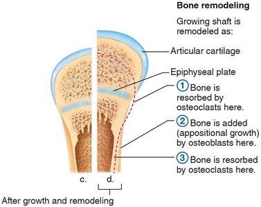
Bone Remodeling
Bone is a dynamic tissue that undergoes continuous remodeling. Osteoclasts resorb bone, while osteoblasts deposit new bone, especially during growth and repair.
Articular cartilage: Maintains smooth joint surfaces.
Epiphyseal plate: Site of longitudinal growth in children.
Osteoclasts: Cells that break down bone matrix.
Osteoblasts: Cells that build new bone matrix.
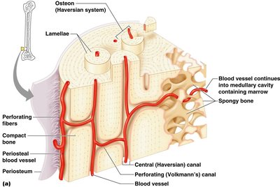
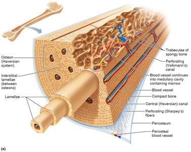
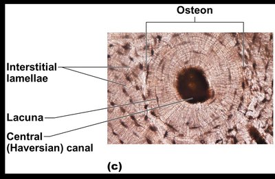
Microscopic Anatomy of Bone
Bone tissue is organized into structural units called osteons (Haversian systems), which contain concentric lamellae, lacunae, and central canals.
Osteon: Fundamental unit of compact bone.
Lamellae: Concentric rings of bone matrix.
Lacunae: Small spaces housing osteocytes.
Central (Haversian) canal: Contains blood vessels and nerves.
Canaliculi: Tiny channels connecting lacunae.
Perforating (Volkmann’s) canal: Transverse canals connecting osteons.
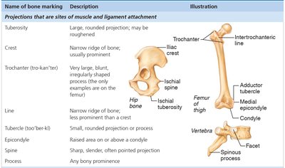
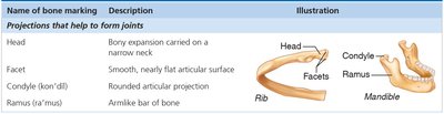
Bone Markings
Surface Features of Bones
Bones have various markings that serve as sites for muscle, tendon, and ligament attachment, or as passages for nerves and blood vessels. These markings are classified as projections (processes) or depressions (cavities).
Projections/processes: Grow out from bone surface (e.g., tuberosity, crest, trochanter).
Depressions/cavities: Indentations in bone (e.g., fossa, foramen).
Name of bone marking | Description | Illustration |
|---|---|---|
Tuberosity | Large, rounded projection; may be roughened | Femur, hip bone |
Crest | Narrow ridge of bone; usually prominent | Hip bone |
Trochanter | Very large, blunt, irregularly shaped process | Femur |
Line | Narrow ridge of bone; less prominent than a crest | Femur |
Tubercle | Small, rounded projection or process | Femur |
Epicondyle | Raised area on or above a condyle | Femur |
Spine | Sharp, slender, often pointed projection | Vertebra |
Process | Any bony prominence | Vertebra |
Name of bone marking | Description | Illustration |
|---|---|---|
Head | Bony expansion carried on a narrow neck | Rib |
Facet | Smooth, nearly flat articular surface | Rib |
Condyle | Rounded articular projection | Mandible |
Ramus | Armlike bar of bone | Mandible |
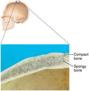
Summary Table: Compact vs. Spongy Bone
Type | Structure | Location | Function |
|---|---|---|---|
Compact bone | Dense, smooth, homogeneous | Diaphysis of long bones, outer layer of flat bones | Support, protection |
Spongy bone | Small needle-like pieces, many open spaces | Epiphyses of long bones, interior of flat bones | Lightens bone, houses marrow |
Additional info:
Bone remodeling is regulated by hormones and mechanical stress.
Bone markings are essential for understanding muscle attachment and joint function.
