 Back
Study Guide: The Skeletal System (Human Anatomy & Physiology)
Study Guide - Smart Notes
 Tailored notes based on your materials, expanded with key definitions, examples, and context.
The Skeletal System
Overview and Components
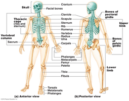
The skeletal system is a complex framework that provides structure, protection, and movement for the human body. It consists of bones, joints, cartilages, and ligaments, and is divided into two main subdivisions: the axial skeleton and the appendicular skeleton.
Bones: The rigid organs forming the skeleton.
Joints: Articulations where two or more bones meet.
Cartilages: Flexible connective tissue found in joints and other areas.
Ligaments: Bands of fibrous tissue connecting bones.
Axial skeleton: Includes the skull, vertebral column, and thoracic cage.
Appendicular skeleton: Includes limbs and girdles (pectoral and pelvic).
Functions of Bones
Key Roles in the Body
Bones serve several essential functions that are critical for overall health and movement.
Support: Provide structural support for the body.
Protection: Shield soft organs (e.g., skull protects brain, rib cage protects thoracic organs).
Movement: Facilitate movement by serving as attachment points for muscles.
Storage: Store minerals (calcium, phosphorus) and fats (yellow marrow).
Blood cell formation: Hematopoiesis occurs in red bone marrow.
Classification of Bones
Types Based on Shape
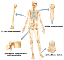
Bones are classified into four groups based on their shape and structure. Each type has distinct characteristics and functions.
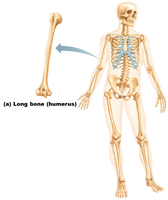
Long bones: Longer than wide, mostly compact bone, found in limbs (e.g., femur, humerus).
Flat bones: Thin, flattened, usually curved, two layers of compact bone with spongy bone in between (e.g., skull, ribs, sternum).
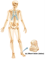
Short bones: Cube-shaped, mostly spongy bone, includes sesamoid bones (e.g., carpals, tarsals, patella).
Irregular bones: Complex shapes, do not fit other categories (e.g., vertebrae, hip bones, facial bones).
Examples of Bone Types
Long bone: Humerus
Flat bone: Sternum
Short bone: Talus
Irregular bone: Vertebra
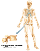
Structure of Bone
Long Bone Anatomy
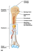
Long bones have a distinct structure, including the diaphysis (shaft), epiphyses (ends), and various membranes and cavities.
Diaphysis: Shaft, composed of compact bone.
Epiphysis: Ends, mostly spongy bone enclosed by compact bone.
Periosteum: Outer covering, fibrous connective tissue.
Endosteum: Lines inner surface of shaft.
Medullary cavity: Contains yellow marrow (fat) in adults, red marrow in children.
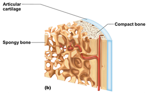
Articular cartilage: Covers epiphyses, reduces friction and absorbs shock.
Epiphyseal line/plate: Site of bone growth; plate is cartilage in children, line is remnant in adults.
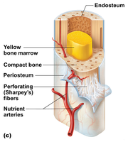
Bone Tissue Types
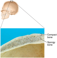
Compact bone: Dense, smooth, homogeneous.
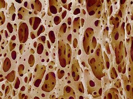
Spongy bone: Small needlelike pieces (trabeculae), many open spaces.
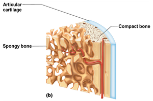
Microscopic Anatomy of Bone
Compact Bone Structure
Compact bone is organized into osteons (Haversian systems), which are the structural and functional units.
Osteocytes: Mature bone cells in lacunae.
Lacunae: Cavities housing osteocytes.
Lamellae: Concentric circles of lacunae around central canal.
Central (Haversian) canal: Contains blood vessels and nerves.
Canaliculi: Tiny canals connecting lacunae to central canal.
Perforating (Volkmann’s) canal: Perpendicular canals carrying blood vessels and nerves.
Spongy Bone Structure
Spongy bone consists of trabeculae and open spaces filled with marrow, blood vessels, and nerves.
Bone Formation, Growth, and Remodeling
Ossification Process
Bone formation (ossification) occurs on hyaline cartilage models or fibrous membranes. Long bone growth involves two major phases:
Osteoblasts cover cartilage model with bone matrix.
Cartilage is digested away, opening up medullary cavity.
By birth, most cartilage is converted to bone except for articular cartilages and epiphyseal plates.
Appositional Growth and Remodeling
Appositional growth: Bones grow in width; osteoblasts add bone matrix to outside, osteoclasts remove from inside.
Remodeling: Occurs throughout life, influenced by calcium levels and mechanical stress.
Calcium regulation: Parathyroid hormone (PTH) activates osteoclasts when blood calcium is low; osteoblasts store calcium when levels are high.
Bone Fractures and Repair
Types of Fractures
Closed (simple) fracture: Does not penetrate skin.
Open (compound) fracture: Penetrates skin.
Fractures are treated by reduction and immobilization. Healing involves four major events:
Hematoma formation
Fibrocartilage callus formation
Bony callus formation
Bone remodeling
Axial Skeleton
Components
The axial skeleton forms the longitudinal axis and includes the skull, vertebral column, and thoracic cage.
Skull: Cranial and facial bones, joined by sutures.
Vertebral column: 26 vertebrae, separated by intervertebral discs.
Thoracic cage: Sternum, ribs, thoracic vertebrae.
Appendicular Skeleton
Components
The appendicular skeleton includes limbs and girdles, facilitating movement and supporting the body.
Pectoral girdle: Clavicle and scapula.
Pelvic girdle: Coxal bones, sacrum, coccyx.
Upper limbs: Humerus, radius, ulna, carpals, metacarpals, phalanges.
Lower limbs: Femur, tibia, fibula, tarsals, metatarsals, phalanges.
Joints (Articulations)
Classification and Structure
Joints are classified functionally (by movement) and structurally (by material and presence of cavity).
Functional: Synarthroses (immovable), amphiarthroses (slightly movable), diarthroses (freely movable).
Structural: Fibrous (immovable), cartilaginous (immovable/slightly movable), synovial (freely movable).
Synovial Joints
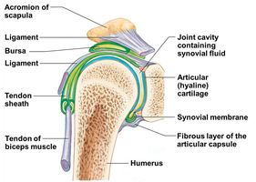
Synovial joints are characterized by a joint cavity containing synovial fluid, articular cartilage, and reinforcing ligaments.
Bursae: Fluid-filled sacs reducing friction.
Tendon sheath: Elongated bursa wrapping around a tendon.
Developmental Aspects of the Skeleton
Growth and Aging
Most fetal skeletal structures are hyaline cartilage or fibrous membranes. Ossification centers develop as the fetus grows. Fontanels (soft spots) allow for skull compression and brain growth, ossifying by age 2.
Growth of cranium relates to brain growth.
Facial skeleton grows with tooth development and respiratory passage enlargement.
Puberty causes female pelvis to broaden and male skeleton to become more robust.
Epiphyseal plates ossify by end of adolescence.
Osteoporosis
Osteoporosis is a bone-thinning disease common in older adults, especially women. It increases fracture risk and can cause vertebral collapse (kyphosis).
Estrogen: Maintains bone density in females.
