 Back
BackStudy Guide: The Skeletal System in Human Anatomy and Physiology
Study Guide - Smart Notes
 Tailored notes based on your materials, expanded with key definitions, examples, and context.
The Skeletal System
Structural Divisions of the Skeleton
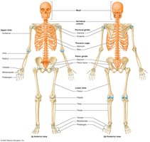
The human skeleton is divided into two main structural divisions: the axial skeleton and the appendicular skeleton. Each division contains specific groups of bones that serve distinct functions in support, movement, and protection.
Axial Skeleton: Forms the longitudinal axis of the body and includes the skull, vertebral column, and thoracic cage.
Appendicular Skeleton: Comprises the bones of the girdles (pectoral and pelvic) and the upper and lower limbs.
Classification of Bones by Shape
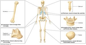
Bones are classified based on their shapes, which relate to their functions and locations in the body.
Long Bones: Longer than they are wide; found in limbs (arms, legs, hands, feet, fingers, toes).
Short Bones: Cube-shaped, small and thick; found in wrists and ankles.
Flat Bones: Thin and broad with parallel surfaces; found in skull, pelvis, sternum, and ribs.
Irregular Bones: Complex shapes; found in vertebrae and pelvic bones.
Sesamoid Bones: Small, flat, oval-shaped; located within tendons (e.g., kneecap).
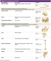
Bone Markings
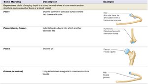
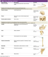
Bone markings are anatomical features on bones that serve as sites for muscle attachment, passage of nerves and blood vessels, and articulation with other bones. They are categorized as depressions, openings, and projections.
Depressions
Facet: Shallow convex or concave surface where two bones articulate.
Fossa: Indentation in a bone into which another structure fits.
Groove: Long indentation along which a narrow structure travels.
Openings
Canal: Tunnel through a bone.
Foramen: Hole in a bone.
Projections
Condyle: Rounded end of a bone that articulates with another bone.
Epicondyle: Small projection usually proximal to a condyle.
Process: Prominent bony projection.
Line: Narrow ridge.
Skull Structure
The skull is composed of cranial and facial bones, united by immovable joints called sutures. These bones protect the brain and support facial structures.
Cranial Bones: Include the frontal, parietal, temporal, occipital, sphenoid, and ethmoid bones.
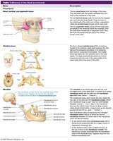
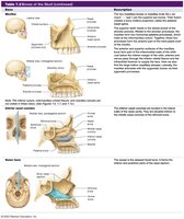
Facial Bones: Include the nasal, maxillae, zygomatic, mandible, lacrimal, palatine, inferior nasal concha, and vomer bones.
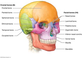
Table: Cranial Bones
Bone | Description |
|---|---|
Frontal | Forms the forehead and part of the eye socket. |
Parietal | Forms the superior and lateral aspects of the skull. |
Occipital | Forms the posterior and base of the skull. |
Temporal | Forms the lateral sides and base of the skull. |
Sphenoid | Forms part of the base and sides of the skull. |
Ethmoid | Forms part of the nasal cavity and eye socket. |
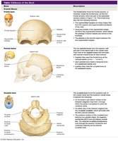
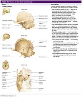
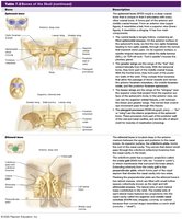
Table: Facial Bones
Bone | Description |
|---|---|
Nasal | Forms the bridge of the nose. |
Maxillae | Forms the upper jaw and part of the eye socket. |
Zygomatic | Forms the cheekbones. |
Mandible | Forms the lower jaw. |
Lacrimal | Forms part of the medial wall of the eye socket. |
Palatine | Forms part of the hard palate. |
Inferior nasal concha | Forms part of the lateral wall of the nasal cavity. |
Vomer | Forms part of the nasal septum. |
Vertebral Column
The vertebral column, or spine, is composed of 33 vertebrae grouped into five regions. It provides structural support, protects the spinal cord, and allows flexibility.
Cervical (7): Located in the neck.
Thoracic (12): Articulate with the ribs.
Lumbar (5): Located in the lower back.
Sacral (5 fused): Articulate with the pelvic bones.
Coccygeal (3–5 fused): Located at the most inferior end.
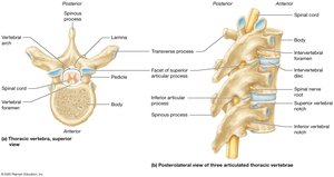
Structure of Vertebrae
Body: Weight-bearing portion.
Vertebral Foramen: Space for the spinal cord.
Pedicles and Laminae: Form the vertebral arch.
Articular Facets: Form joints between vertebrae.
Transverse Processes: Lateral projections for muscle attachment.
Spinous Process: Posterior projection for muscle attachment.
Cervical Vertebrae
Smallest vertebrae with transverse foramina.
Atlas (C1): No vertebral body or spinous process.
Axis (C2): Contains the dens (odontoid process).
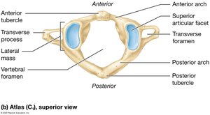
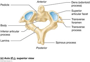
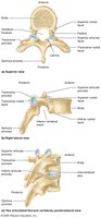
Thoracic Vertebrae
Articulate with ribs and have long spinous processes.
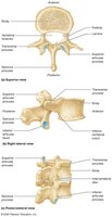
Lumbar Vertebrae
Largest vertebrae with thick bodies and short spinous processes.
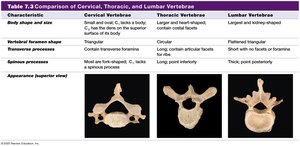
Comparison Table: Cervical, Thoracic, and Lumbar Vertebrae
Characteristic | Cervical | Thoracic | Lumbar |
|---|---|---|---|
Body shape | Small, oval | Larger, heart-shaped | Largest, kidney-shaped |
Transverse foramen | Present | Absent | Absent |
Spinous process | Forked | Long, point inferiorly | Thick, point posteriorly |
Thoracic Cage
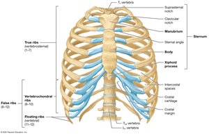
The thoracic cage protects vital organs and supports the upper body. It consists of the sternum and 12 pairs of ribs.
Sternum: Manubrium, body, xiphoid process.
Ribs:
True ribs (1–7): Attach directly to the sternum.
False ribs (8–12): Do not attach directly.
Floating ribs (11–12): Do not attach to the sternum.
Pectoral Girdle and Upper Limb
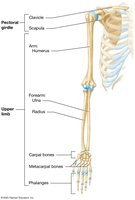
The pectoral girdle consists of the clavicle and scapula, which anchor the upper limb to the trunk. The upper limb includes the humerus, radius, ulna, carpals, metacarpals, and phalanges.
Clavicle: S-shaped bone connecting the sternum and scapula.
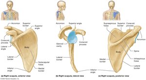
Scapula: Flat bone with several important features (glenoid cavity, acromion, coracoid process).
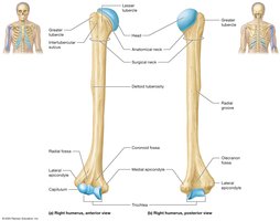
Humerus: Upper arm bone; articulates with scapula and forearm bones.
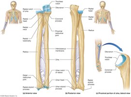
Radius and Ulna: Forearm bones; radius is lateral, ulna is medial.
Pelvic Girdle and Lower Limb
The pelvic girdle consists of the sacrum and pelvic bones (ilium, ischium, pubis). The lower limb includes the femur, patella, tibia, fibula, tarsals, metatarsals, and phalanges.
Pelvic Bone: Three fused bones (ilium, ischium, pubis) form the acetabulum and obturator foramen.
Femur: Thigh bone; articulates with the pelvis and tibia.
Patella: Sesamoid bone in the quadriceps tendon.
Tibia: Medial leg bone; forms the medial malleolus.
Fibula: Lateral leg bone; forms the lateral malleolus.
Tarsals: Ankle bones; include talus and calcaneus.
Metatarsals and Phalanges: Bones of the foot and toes.
Summary Table: Major Bones and Their Functions
Bone | Function |
|---|---|
Skull | Protects brain, supports facial structures |
Vertebral column | Supports body, protects spinal cord |
Thoracic cage | Protects heart and lungs |
Pectoral girdle | Anchors upper limb |
Pelvic girdle | Anchors lower limb, supports body weight |
Upper limb bones | Movement and manipulation |
Lower limb bones | Support and locomotion |
Additional info: The notes above expand on brief points with academic context, definitions, and examples to ensure completeness and clarity for college-level study.
