 Back
BackStudy Guide: The Special Senses in Human Anatomy & Physiology
Study Guide - Smart Notes
 Tailored notes based on your materials, expanded with key definitions, examples, and context.
Chapter 15: The Special Senses
Overview of the Special Senses
The special senses are distinguished from general senses by their reliance on specialized sensory organs located in discrete regions of the head. These senses include smell (olfaction), taste (gustation), vision, hearing (audition), and vestibular sensation (balance). Each sense detects specific stimuli and transduces them into neural signals for interpretation by the brain.
Comparison of General and Special Senses
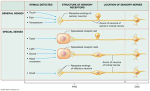
General senses detect stimuli such as touch, pain, and temperature, while special senses detect specific stimuli like light, sound, head movement, and chemicals. The structure and location of sensory receptors differ between these two categories.
General Senses: Receptive ends of sensory neurons; transmitted via spinal and cranial nerves.
Special Senses: Specialized receptor cells (except olfaction, which uses neurons); transmitted via cranial nerves, all housed in the head.
Sensory Transduction
Transduction is the process by which a physical or chemical stimulus is converted into an action potential. Special senses transduce environmental stimuli (e.g., light, sound, chemicals) into neural signals, which are processed by sensory nuclei, thalamus, and primary cortical areas, then integrated in association areas.
Olfaction (Smell)
Structures of Olfaction
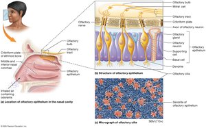
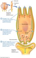
Olfaction enables detection of odorants in the air. The olfactory epithelium is located in the superior region of each nasal cavity and contains three cell types:
Olfactory Neurons: Modified bipolar neurons with olfactory cilia; axons form the olfactory nerve, synapse on mitral cells in the olfactory bulb, and travel through the olfactory tract.
Basal Cells: Stem cells that replace olfactory neurons (lifespan: 30–60 days).
Supporting Cells: Columnar cells that support and surround olfactory neurons.
Physiology of Olfaction
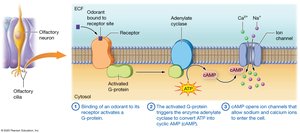
Odorants bind to odorant-binding proteins, activating a G-protein, which triggers adenylate cyclase to convert ATP into cAMP. cAMP opens ion channels, allowing Na+ and Ca2+ to enter, depolarizing the membrane.
Humans: ~350 genes for receptor proteins; each can bind multiple odorants.
Olfactory Pathway: Olfactory nerve → olfactory bulb → primary olfactory cortex (temporal lobe); only sensory pathway without thalamic synapse.
Limbic System: Emotional and visceral responses to odors.
Gustation (Taste)
Structures of Gustation: Taste Buds
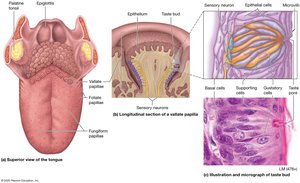
Taste buds are clusters of receptor and supporting cells on the tongue and oral cavity. The tongue is covered with papillae:
Vallate Papillae: Large, dome-shaped, hundreds of taste buds.
Fungiform Papillae: Mushroom-shaped, few taste buds.
Foliate Papillae: Ridges, taste buds only in childhood.
Filiform Papillae: No taste buds, detect texture and temperature.
Taste bud cell types:
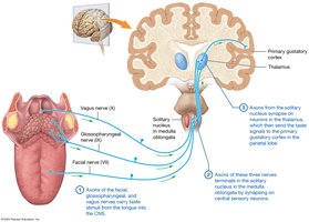
Gustatory Cells: Specialized epithelial cells with microvilli; synapse with sensory neurons (facial, glossopharyngeal, vagus nerves).
Basal Cells: Stem cells for gustatory cell replacement.
Supporting Cells: Physical support.
Physiology of Gustation
Taste sensations depend on chemical detection and receptor activation:
Sweet: Simple sugars (glucose, fructose).
Sour: Hydrogen ions (e.g., citric acid).
Salty: Metal ions (Na+, K+).
Bitter: Alkaloids (coffee, rancid foods); protective sensitivity.
Umami: Glutamate or amino acids.
Activation of taste receptors involves ion movement, depolarization, opening of voltage-gated Ca2+ channels, and neurotransmitter release.
The Gustatory Pathway
Taste stimuli are carried by facial, glossopharyngeal, and vagus nerves to the solitary nucleus in the medulla, then to the thalamus, and finally to the primary gustatory cortex in the parietal lobe. Integration with visual and olfactory stimuli occurs in the insula and inferior frontal lobe.
Vision
Accessory Structures of the Eye
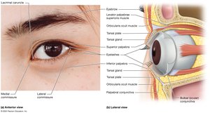
Accessory structures support, protect, and move the eyeball:
Eyelids: Cover the orbit, distribute tears, prevent foreign objects.
Eyebrows: Prevent perspiration, reduce glare, aid expression.
Eyelashes: Trigger blink reflex.
Conjunctiva: Epithelial membrane; lines eyelids and covers eye.
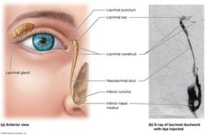
Lacrimal Apparatus
Produces and drains tears, lubricating and cleaning the eye. Tears flow from the lacrimal gland to the lacrimal puncta, canaliculi, sac, and nasolacrimal duct.
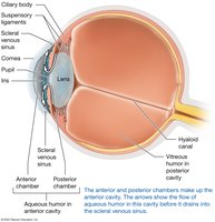
The Eyeball: Structure and Layers
The eyeball is a hollow sphere with three tissue layers:
Fibrous Layer: Sclera (white, collagenous), cornea (translucent, allows light entry).
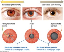
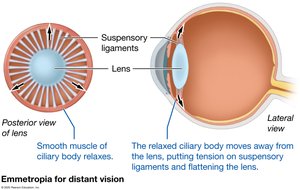
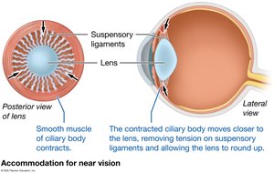
Vascular Layer: Choroid (blood supply, pigment), ciliary body (smooth muscle, lens focus), iris (pigmented, controls pupil size).
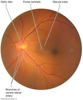
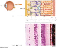
Neural Layer (Retina): Pigmented layer (reduces light scatter), photoreceptors (rods and cones), macula lutea (high acuity), optic disc (blind spot).
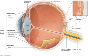
Lens and Eye Chambers
The lens focuses light on the retina. The posterior cavity contains vitreous humor, maintaining eyeball shape. The anterior cavity is divided into anterior and posterior chambers, filled with aqueous humor, which drains via the scleral venous sinus.
Principles of Light and Vision
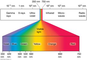
Electromagnetic Spectrum and Visible Light
Vision is the perception of light, a form of electromagnetic radiation. The visible spectrum ranges from ~350 nm (violet) to ~750 nm (red). Photons stimulate retinal photoreceptors.
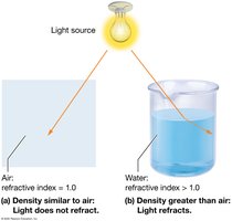
Refraction and Lenses
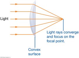
Light bends (refracts) when passing through materials with different refractive indices. The cornea and lens focus light on the retina. Convex lenses converge light; concave lenses diverge it.
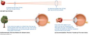
Focusing Light on the Retina
Light from distant objects requires little refraction; the lens remains flattened (emmetropic state). Near objects require accommodation: the ciliary body contracts, suspensory ligaments loosen, and the lens rounds up for greater refraction.
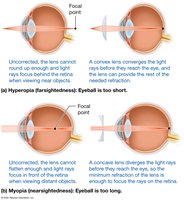
Errors of Refraction
Condition | Cause | Correction |
|---|---|---|
Presbyopia | Stiff lens, reduced accommodation | Reading glasses/bifocals |
Hyperopia (Farsightedness) | Eyeball too short/cornea too flat | Convex lens |
Myopia (Nearsightedness) | Eyeball too long/cornea too curved | Concave lens |
Astigmatism | Irregular lens/cornea curvature | Corrective lenses/LASIK |
Photoreceptors and the Retina
Retinal Structure and Cell Types
Photoreceptors (rods and cones) synapse with bipolar cells, which communicate with retinal ganglion cells. Horizontal and amacrine cells modulate transmission and respond to light changes.
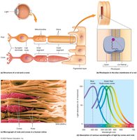
Rods and Cones
Cones: Color, high-acuity vision, require bright light, concentrated in fovea.
Rods: Black/white, low-acuity vision, sensitive in dim light, peripheral vision.
Color Blindness
Defective gene for cone pigments, most commonly red-green. Sex-linked disorder, more common in males. Ishihara plates are used for diagnosis.

Transduction in Photoreceptors
Light triggers conversion of cis-retinal to trans-retinal, activating transducin and phosphodiesterase, closing sodium channels, and hyperpolarizing the cell. In the dark, cells are depolarized and release neurotransmitters.
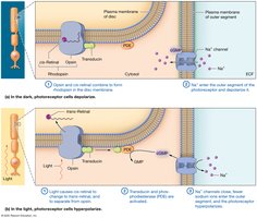
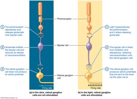
Image Processing by the Retina
In the dark, photoreceptors release glutamate, inhibiting bipolar cells and preventing action potentials in ganglion cells. In the light, hyperpolarization frees bipolar cells, which depolarize and stimulate ganglion cells to send action potentials to the brain.
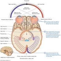
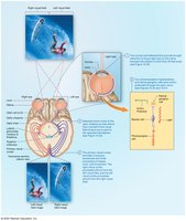
The Visual Pathway
Structures and Processing
Visual stimuli are detected by the retina, processed by the optic nerve, optic chiasma, optic tracts, thalamus, and primary visual cortex. Binocular vision enables depth perception; the brain integrates images for motion, shape, and color.
Anatomy of the Ear
Regions of the Ear
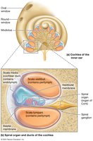
The ear consists of three regions: outer (auricle, external auditory canal, tympanic membrane), middle (auditory ossicles, pharyngotympanic tube), and inner (bony and membranous labyrinth, cochlea, vestibule, semicircular canals).
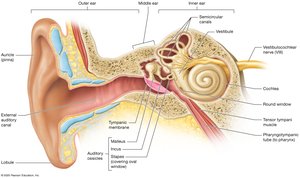
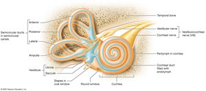
Inner Ear Structure
The inner ear contains the vestibule (utricle, saccule), semicircular canals (ducts), and cochlea (scala vestibuli, scala tympani, cochlear duct, spiral organ).
Principles of Sound
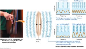
Sound Waves
Sound waves are produced by displacement of air molecules. Pitch (frequency) is measured in hertz (Hz), and loudness (amplitude) in decibels (dB).
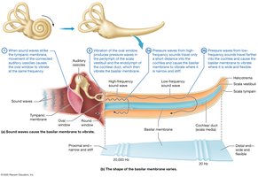
Transmission of Sound to the Inner Ear
Sound waves strike the tympanic membrane, move ossicles, vibrate the oval window, and produce pressure waves in perilymph and endolymph, vibrating the basilar membrane. High-frequency sounds vibrate the narrow, stiff region; low-frequency sounds vibrate the wide, flexible region.
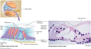
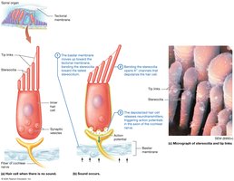
Processing of Sound in the Inner Ear
The spiral organ contains hair cells with stereocilia. Bending stereocilia opens potassium channels, depolarizing the cell and releasing neurotransmitters, triggering action potentials in the cochlear nerve.
Vestibular Sensation
Static and Dynamic Equilibrium
Equilibrium depends on vision, proprioceptors, and the vestibular system. Static equilibrium is monitored by the utricle and saccule; dynamic equilibrium by semicircular ducts.
The Vestibular Sensation Pathway
Action potentials propagate to vestibular nuclei, thalamus, cranial nerve nuclei, cerebellum, and spinal cord, coordinating posture, awareness, and eye movements.
Integration of Special Senses
Special senses integrate with other brain areas to form a coherent perception. Receptors detect stimuli, cranial nerves transmit signals, thalamus processes (except olfaction), primary cortices generate awareness, and the frontal lobe and limbic system integrate experiences.
