 Back
BackStudy Guide: The Special Senses in Human Anatomy & Physiology
Study Guide - Smart Notes
 Tailored notes based on your materials, expanded with key definitions, examples, and context.
The Special Senses
Overview of the Special Senses
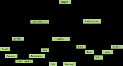
The special senses include vision, hearing, equilibrium, taste, and smell. These senses are distinguished from general senses by their specialized receptor cells and complex neural pathways. Special senses allow humans to interact with and interpret their environment in highly specific ways.
General Senses: Touch, pain, temperature, pressure, proprioception.
Special Senses: Smell (olfaction), taste (gustation), vision, hearing, balance (equilibrium).
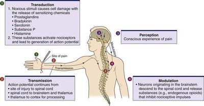
Sensory Transduction: The process by which a physical or chemical stimulus is converted into an action potential that can be interpreted by the brain.
Pain and Pain Perception
Pain is an unpleasant sensation associated with actual or potential tissue damage. It serves as a protective mechanism, motivating avoidance of harm. Pain can be classified based on its origin and characteristics.
Types of Pain:
Visceral pain: Originates from internal organs.
Deep somatic pain: From bones, joints, muscles.
Superficial somatic pain: From skin.
Neuropathic pain: From nerve injuries, often described as stabbing, burning, or tingling.
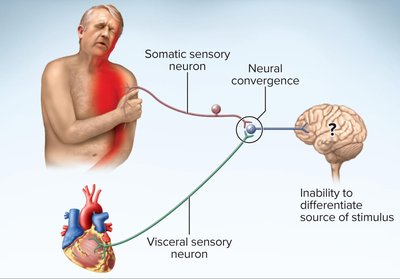
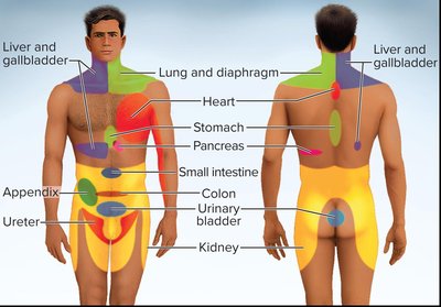
Referred Pain: Pain felt in a location different from its source due to neural convergence. Example: Left arm pain during a heart attack.
Phantom Pain
Phantom pain is the sensation of pain perceived in a body part that has been amputated. It is a real phenomenon originating from the spinal cord and brain, not a psychological issue.
Theories for Cause: Mixed signals from remaining nerves, sensory remapping, neuromas, trauma, physical memory, and changes in neurosignature.
Example: Post-amputation pain from a missing limb.

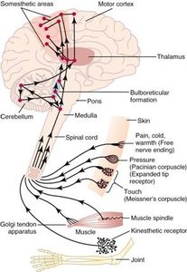
Somatosensory Projection Pathways
Somatosensory pathways transmit sensory information from the body to the brain. These pathways involve multiple neurons and relay stations.
First-order neuron: Carries information via cranial or spinal nerves.
Second-order neuron: Crosses to the opposite side of the CNS and ends in the thalamus (except proprioception, which ends in the cerebellum).
Third-order neuron: Projects from the thalamus to the primary cerebral cortex.
Olfaction (Smell)
Structures and Physiology of Olfaction
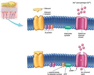
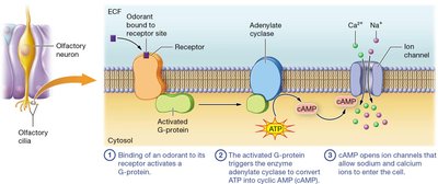
Olfaction is the sense of smell, mediated by olfactory receptors located in the nasal cavity. Odorants dissolve in mucus and bind to receptors on olfactory hairs (cilia), initiating action potentials.
Olfactory Epithelium: Contains bipolar olfactory neurons, supporting cells, and basal cells.
Odorant Activation: Odorants bind to membrane receptors, causing depolarization and action potential generation.
Threshold: Humans can detect about 400,000 odorants; sensitivity is extremely high.
Adaptation: Receptor saturation leads to decreased sensitivity (nose-blindness).
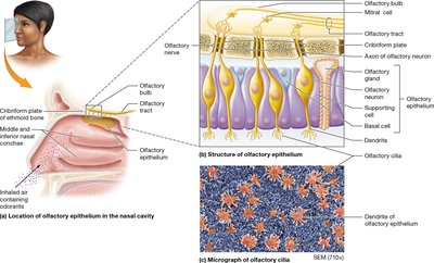
Olfactory Neuronal Pathways
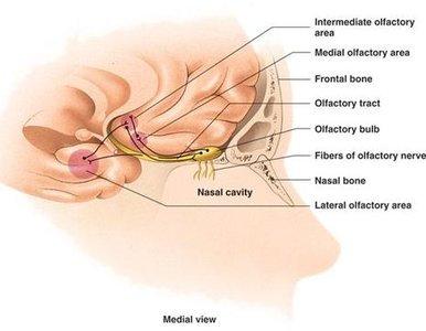
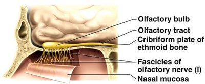
Olfactory neurons pass through the cribriform plate to the olfactory bulbs, synapse with mitral cells, and project to the olfactory cortex. Unlike other senses, olfactory signals bypass the thalamus initially.
Pathway: Olfactory neurons → olfactory bulb → olfactory tract → olfactory cortex (inferomedial temporal lobe and inferior frontal lobe).
Limbic System: Olfactory signals evoke emotional and visceral responses via connections to the amygdala, hippocampus, and hypothalamus.
Frontal Lobe Regions:
Lateral olfactory area: Conscious perception
Medial olfactory area: Emotional/visceral reactions
Intermediate olfactory area: Modifies incoming information
Gustation (Taste)
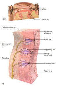
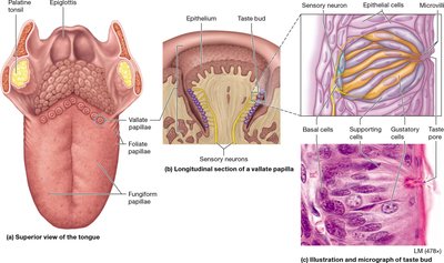
Structure and Function of Taste Buds
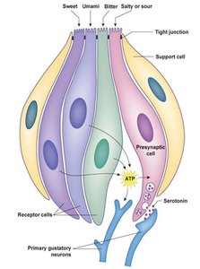
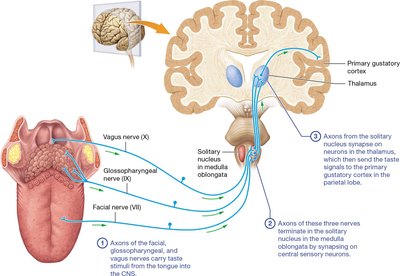
Gustation is the sense of taste, mediated by taste buds located primarily on papillae of the tongue. Each taste bud contains gustatory cells with microvilli (gustatory hairs) that extend into taste pores.
Taste Buds: 50-150 gustatory cells per bud; each cell is sensitive to one tastant.
Perception: Influenced by texture, temperature, and olfaction.
Adaptation: Rapid at both the taste bud and CNS level.
Thresholds: Bitter taste has the lowest threshold (highest sensitivity).
Primary Taste Sensations
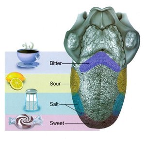
There are five primary taste sensations: sour, salty, bitter, sweet, and umami. Each is detected by specific receptors distributed across the tongue and oral cavity.
Sour: Lateral aspects of tongue; most sensitive.
Salty: Tip of tongue; shares lowest sensitivity with sweet.
Bitter: Posterior aspect; highest sensitivity, often to alkaloids.
Sweet: Tip of tongue; shares lowest sensitivity with salty.
Umami: Scattered sensitivity; caused by amino acids (glutamate).
Taste Receptor Activation and Gustatory Pathway
Tastants must dissolve in saliva to enter the taste pore and activate gustatory cells. The basal end of each cell synapses with a primary sensory neuron. Taste information is relayed via cranial nerves VII (facial), IX (glossopharyngeal), and X (vagus) to the brain.
Pathway:
Anterior 2/3 tongue: Facial nerve (VII)
Posterior 1/3 tongue: Glossopharyngeal nerve (IX)
Superior pharynx and epiglottis: Vagus nerve (X)
Integration: Signals are relayed to the insula, medulla oblongata, thalamus, and taste area of cortex. Emotional responses are produced via the limbic system.
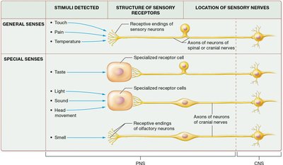
Summary Table: General vs Special Senses
Type of Sense | Stimuli Detected | Structure of Sensory Receptors | Location of Sensory Nerves |
|---|---|---|---|
General Senses | Touch, Pain, Temperature | Receptive endings of sensory neurons | Axons of spinal or cranial nerves |
Special Senses | Taste, Light, Sound, Head movement, Smell | Specialized receptor cells (except olfaction: olfactory neurons) | Axons of cranial nerves |
Integration of Special Senses
Frontal Lobe and Limbic System
The frontal lobe and limbic system integrate signals from the special senses, creating a meaningful perception of the environment. This integration allows for emotional and visceral responses to sensory stimuli.
Frontal Lobe: Conscious perception and interpretation.
Limbic System: Emotional and visceral reactions.
Additional info: The special senses are essential for survival, communication, and interaction with the environment. Their pathways are highly specialized and involve complex neural integration.
