 Back
BackStudy Guide: The Spinal Cord, Spinal Nerves, and Spinal Reflexes
Study Guide - Smart Notes
 Tailored notes based on your materials, expanded with key definitions, examples, and context.
The Nervous System
Structural and Functional Organization
The nervous system is divided into the central nervous system (CNS) and peripheral nervous system (PNS), each with distinct structural and functional roles. - Central Nervous System (CNS): Consists of the brain and spinal cord, serving as processing centers for sensory input and motor output. - Peripheral Nervous System (PNS): Composed of cranial and spinal nerves, responsible for transmitting sensory information to the CNS and carrying motor commands to effectors. - Reflexes: Quick, automatic responses to stimuli; spinal reflexes are controlled by the spinal cord without brain input. 
The Spinal Cord
Structure and Functions
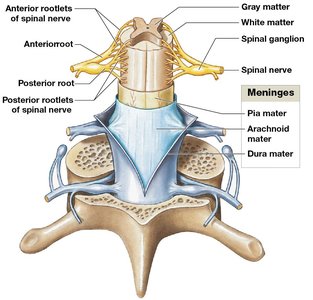
The spinal cord is a major organ of nervous tissue housed within protective membranes (meninges) and the vertebral column. It carries sensory and motor information between the brain and most parts of the body and is the origin of spinal nerves. - Length: Approximately 45 cm (18 in.) long, 14 mm (0.55 in.) wide. - Regions: Cervical, thoracic, lumbar, and sacral. - Gray Matter: Contains neuronal cell bodies. - White Matter: Contains myelinated axons.
Gross Anatomy and Segmentation
- 31 segments: Each gives rise to a pair of spinal nerves. - Bilateral symmetry: Grooves divide the cord into left and right. - Posterior median sulcus: Shallow groove on the posterior side. - Anterior median fissure: Deeper groove on the anterior side. - Central canal: Internal passageway containing cerebrospinal fluid (CSF). 
Enlargements and Distal End
- Cervical enlargement: Supplies nerves to the shoulder and upper limb. - Lumbosacral enlargement: Supplies nerves to the pelvis and lower limb. - Conus medullaris: Tapered end below lumbar enlargement. - Cauda equina: Nerve roots extending below conus medullaris. - Filum terminale: Fibrous tissue thread attaching to coccygeal ligament.
Spinal Roots and Ganglia
- Anterior root (ventral root): Motor neuron axons. - Posterior root (dorsal root): Sensory neuron axons. - Spinal ganglia (dorsal root ganglia): Cell bodies of sensory neurons. 
Spinal Nerves
- Mixed nerves: Contain both afferent (sensory) and efferent (motor) fibers. - Branches: White ramus communicans (myelinated), gray ramus communicans (unmyelinated), posterior ramus (back), anterior ramus (trunk and limbs).
Naming Spinal Nerves
- Designation: By vertebral region and number (e.g., C1, T1).
Spinal Meninges
Specialized membranes provide stability, shock absorption, and blood supply pathways. - Dura mater: Tough, outer layer. - Arachnoid mater: Middle layer, includes arachnoid membrane and trabeculae. - Pia mater: Inner layer, firmly attached to neural tissue. - Meningitis: Infection of meninges. 

Gray Matter and White Matter
Roles in Processing and Relaying Information
- Gray matter: Cell bodies, neuroglia, unmyelinated axons; integrates information and initiates commands. - White matter: Myelinated and unmyelinated axons; carries information between regions.
Organization of Gray Matter
- Nuclei: Masses of gray matter organized into horns. - Posterior horns: Somatic and visceral sensory nuclei. - Anterior horns: Somatic motor nuclei. - Lateral horns: Visceral motor nuclei (thoracic/lumbar). - Gray commissure: Band around central canal. 
Organization of White Matter
- Columns: Posterior, anterior, and lateral. - Tracts: Bundles of axons relaying information. - Ascending tracts: Sensory information to the brain. - Descending tracts: Motor commands to the spinal cord. 
Spinal Nerves and Plexuses
Connective Tissue Layers
Spinal nerves are surrounded by three connective tissue layers:
Layer | Description |
|---|---|
Epineurium | Outermost; network of collagen fibers |
Perineurium | Middle; separates nerve into fascicles |
Endoneurium | Innermost; surrounds individual axons |

Dermatomes and Peripheral Neuropathies
- Dermatome: Region of skin monitored by a single pair of spinal nerves. - Clinical importance: Loss of sensation indicates nerve damage. - Peripheral neuropathies: Regional losses of function due to trauma/compression. - Shingles: Varicella-zoster virus attacks spinal nerves, causing rash along dermatomes. 

Nerve Plexuses
Complex networks formed from anterior rami of adjacent spinal nerves, allowing multiple nerves to supply the same structures.
Plexus | Spinal Nerves | Major Nerves |
|---|---|---|
Cervical | C1–C5 | Phrenic, lesser occipital, great auricular, transverse cervical, supraclaviculars, ansa cervicalis |
Brachial | C5–T1 | Musculocutaneous, radial, median, ulnar |
Lumbar | T12–L4 | Iliohypogastric, ilio-inguinal, genitofemoral, lateral femoral cutaneous, femoral, obturator |
Sacral | L4–S4 | Superior/inferior gluteal, posterior femoral cutaneous, sciatic (fibular, tibial), pudendal |

Neuronal Pools and Neural Circuits
Functional Organization
Neuronal pools are groups of interconnected interneurons with limited input and output. They may stimulate or depress regions of the CNS.
Patterns of Neural Circuits
- Divergence: One neuron spreads information to many. - Convergence: Several neurons synapse on a single neuron. - Serial processing: Information moves sequentially. - Parallel processing: Several neurons process the same information simultaneously. - Reverberation: Positive feedback loop maintains activity.
Reflexes
Neural Reflexes and Reflex Arcs
Reflexes are rapid, automatic responses to stimuli, preserving homeostasis. The reflex arc is the route nerve impulses follow to produce a reflex. Steps in a Reflex Arc: 1. Stimulus activates a receptor 2. Activation of a sensory neuron 3. Information processing in CNS 4. Activation of a motor neuron 5. Response by a peripheral effector
Classification of Reflexes
- Development: Innate (inborn) vs. acquired (learned) - Motor response: Somatic (muscle) vs. visceral (internal organs) - Complexity: Monosynaptic (single synapse, fast) vs. polysynaptic (multiple synapses, slower) - Site: Spinal (spinal cord) vs. cranial (brain)
Spinal Reflexes
Monosynaptic Reflexes
- Stretch reflex: Regulates skeletal muscle length; example is the patellar reflex.
Muscle Spindle
Sensory receptors involved in stretch reflexes, made of intrafusal muscle fibers and surrounded by extrafusal fibers.
Polysynaptic Reflexes
- Tendon reflex: Prevents excessive tension in muscles. - Withdrawal reflex: Moves body part away from stimulus; involves reciprocal inhibition. - Crossed extensor reflex: Contralateral response to maintain balance.
General Characteristics of Polysynaptic Reflexes
1. Involve pools of interneurons 2. Involve multiple spinal segments 3. Reciprocal inhibition 4. Reverberating circuits 5. Cooperation among reflexes
Brain Control of Spinal Reflexes
Integration and Modification
Higher brain centers can facilitate, inhibit, or fine-tune spinal reflexes. - Reinforcement: Enhances reflexes via excitatory interneurons. - Inhibition: Suppresses reflexes via inhibitory neurons.
Plantar and Babinski Reflexes
- Plantar reflex: Normal in adults; toe-curling. - Babinski reflex: Normal in infants; toe-fanning. Presence in adults may indicate CNS damage. Example: Stroking the lateral sole of the foot.
Summary Table: Spinal Cord and Reflexes
Feature | Description |
|---|---|
Spinal Cord | Central nervous tissue, origin of spinal nerves |
Spinal Nerves | Mixed nerves, sensory and motor fibers |
Meninges | Dura mater, arachnoid mater, pia mater |
Gray Matter | Cell bodies, integration |
White Matter | Axons, information relay |
Reflex Arc | Stimulus → receptor → sensory neuron → CNS → motor neuron → effector |
Reflex Types | Monosynaptic, polysynaptic, somatic, visceral, spinal, cranial |
