Back
Study Guide: The Spinal Cord, Spinal Nerves, and Spinal Reflexes
Study Guide - Smart Notes
 Tailored notes based on your materials, expanded with key definitions, examples, and context.
The Nervous System
Structural Organization
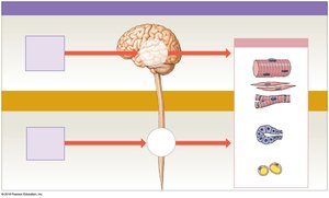
The nervous system is divided into the central nervous system (CNS) and peripheral nervous system (PNS). The CNS consists of the brain and spinal cord, which serve as processing centers. The PNS includes cranial nerves and spinal nerves, responsible for transmitting sensory input and motor responses.
Central Nervous System (CNS): Brain and spinal cord; main processing centers.
Peripheral Nervous System (PNS): Cranial and spinal nerves; pathways for sensory input and motor output.
Reflexes
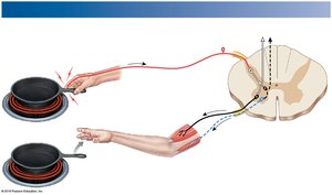
Reflexes are quick, automatic nerve responses triggered by specific stimuli. Spinal reflexes are controlled by the spinal cord alone, without input from the brain. For example, dropping a hot pan is a spinal reflex that occurs before pain is perceived.
Spinal Reflexes: Controlled by the spinal cord; rapid and automatic.
Example: Withdrawal from a hot object.
The Spinal Cord
Gross Anatomy
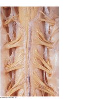
The spinal cord is housed within protective membranes (meninges) and the vertebral column. It carries sensory and motor information between the brain and most other parts of the body and gives rise to spinal nerves.
Length: About 18 inches (45 cm); width 1/2 inch (14 mm).
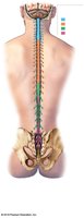
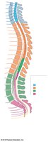
Regions: Cervical, thoracic, lumbar, sacral.
Segments: 31 segments, each giving rise to a pair of spinal nerves.
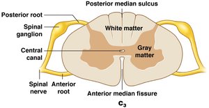
Grooves: Posterior median sulcus (posterior), anterior median fissure (anterior).
Central Canal: Contains cerebrospinal fluid (CSF).
Enlargements and Distal End
Cervical Enlargement: Supplies shoulder and upper limb.
Lumbosacral Enlargement: Supplies pelvis and lower limb.
Conus Medullaris: Tapered, conical end below lumbar enlargement.
Cauda Equina: Nerve roots extending below conus medullaris.
Filum Terminale: Thin thread of fibrous tissue attaching to coccygeal ligament.
Spinal Roots and Ganglia
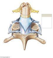
Ventral Root: Anterior root; axons of motor neurons.
Dorsal Root: Posterior root; axons of sensory neurons.
Dorsal Root Ganglia: Contain cell bodies of sensory neurons.
Spinal Nerves
Formation: Union of posterior and anterior roots.
Mixed Nerves: Contain both afferent (sensory) and efferent (motor) fibers.
Rami: Posterior ramus supplies back; anterior ramus supplies body wall, skin, limbs.
Naming Spinal Nerves
Designation: By vertebral region and number (e.g., C1, T1).
The Spinal Meninges
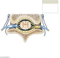
Three specialized membranes surround the spinal cord, providing protection and carrying blood supply.
Dura Mater: Outermost, tough layer.
Arachnoid Mater: Middle, weblike layer.
Pia Mater: Innermost, mesh of collagen and elastic fibers.
Gray Matter and White Matter
Functional Organization
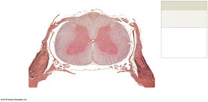
Gray matter consists of cell bodies, neuroglia, and unmyelinated axons. White matter is mostly myelinated axons.
Gray Matter: Organized into nuclei and horns (posterior, anterior, lateral).
White Matter: Organized into columns (posterior, anterior, lateral).
Tracts
Ascending Tracts: Carry sensory information to the brain.
Descending Tracts: Carry motor commands to the spinal cord.
Spinal Nerves and Plexuses
Connective Tissue Layers
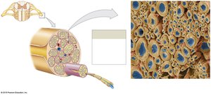
Epineurium: Outermost layer.
Perineurium: Middle layer; separates nerve into fascicles.
Endoneurium: Innermost; surrounds individual axons.
Dermatomes
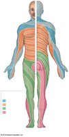
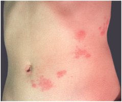
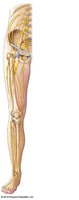
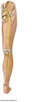
A dermatome is a specific bilateral region of skin supplied by a single pair of spinal nerves. Mapping dermatomes helps diagnose nerve damage.
Peripheral Neuropathies: Regional losses of neural function.
Shingles: Rash occurs along dermatomes.
Nerve Plexuses
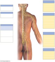
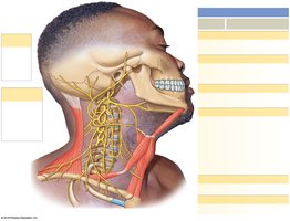
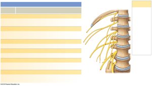
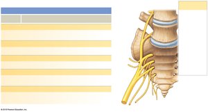
Nerve plexuses are complex networks formed from blended fibers of anterior rami of adjacent spinal nerves. They allow multiple spinal nerves to supply the same structures.
Cervical Plexus: C1–C5; innervates neck and diaphragm.
Brachial Plexus: C5–T1; innervates pectoral girdle and upper limb.
Lumbar Plexus: T12–L4; innervates abdominal muscles and lower extremities.
Sacral Plexus: L4–S4; innervates pelvic girdle and lower limb.
Neuronal Pools
Functional Organization
Sensory Neurons: Bring information into CNS.
Interneurons: Interpret, plan, and coordinate signals.
Motor Neurons: Send commands from CNS to effectors.
Patterns of Neural Circuits
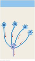
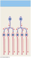
Divergence: Spreads information from one neuron to many.
Convergence: Several neurons synapse on a single neuron.
Serial Processing: Information moves sequentially.
Parallel Processing: Several neurons process information simultaneously.
Reverberation: Positive feedback loop; continues stimulation.
Reflexes
Neural Reflexes and Reflex Arc
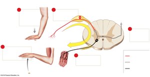
Neural reflexes are rapid, automatic responses to specific stimuli. The reflex arc consists of five components: sensory receptor, sensory neuron, information processing in CNS, motor neuron, and effector.
Reflex Arc Steps: 1. Stimulus activates receptor; 2. Action potential in sensory neuron; 3. Information processing in CNS; 4. Motor neuron activation; 5. Effector response.
Classification of Reflexes
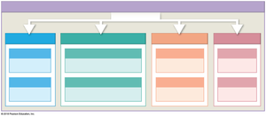
Development: Innate (inborn) or acquired (learned).
Motor Response: Somatic (skeletal muscle) or visceral (autonomic).
Complexity: Monosynaptic (one synapse) or polysynaptic (multiple synapses).
Processing Site: Spinal or cranial.
Spinal Reflexes
Monosynaptic Reflexes
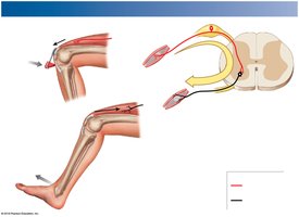
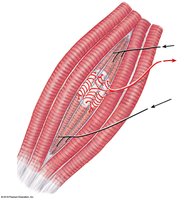
Monosynaptic reflexes involve a single synapse between sensory and motor neurons. The stretch reflex regulates skeletal muscle length and is very rapid.
Example: Patellar (knee-jerk) reflex.
Muscle Spindle Structure
Muscle Spindle: Receptor in stretch reflex; made of intrafusal fibers.
Gamma Motor Neurons: Innervate intrafusal fibers.
Extrafusal Fibers: Typical skeletal muscle fibers surrounding spindle.
Postural Reflexes
Function: Maintain upright posture; involve both monosynaptic and polysynaptic reflexes.
Polysynaptic Reflexes
Tendon Reflex: Prevents excessive tension; involves Golgi tendon organs.
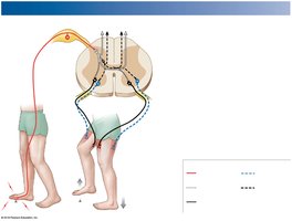
Withdrawal Reflex: Moves body part away from stimulus; involves reciprocal inhibition.
Crossed Extensor Reflex: Coordinates with flexor reflex; involves contralateral reflex arcs.
Characteristics of Polysynaptic Reflexes
Involve pools of interneurons
May cause excitation or inhibition
Involve more than one spinal segment
Can activate muscles in multiple areas
Involve reciprocal inhibition
Have reverberating circuits
Several reflexes may cooperate for coordinated response
The Brain Can Alter Spinal Reflexes
Integration and Control
Brain Centers: Can facilitate or inhibit spinal reflex motor patterns.
Voluntary Movements: Brain can activate motor patterns through descending pathways.
Reinforcement: Higher centers enhance reflexes by stimulating excitatory interneurons.
Inhibition: Higher centers inhibit reflexes by stimulating inhibitory neurons.
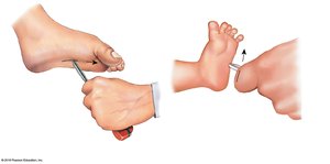
Plantar Reflex and Babinski Reflex
Plantar Reflex: Normal in adults; toe-curling response.
Babinski Reflex: Normal in infants; pathological in adults.