 Back
Chapter 13 study guide
Study Guide - Smart Notes
 Tailored notes based on your materials, expanded with key definitions, examples, and context.
The Spinal Cord: Anatomy and Protective Coverings
Location and Protection
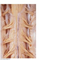
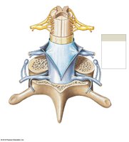
The spinal cord is a vital structure of the central nervous system, enclosed within the vertebral column and surrounded by protective meninges. These coverings provide both physical protection and a supportive environment for neural function.
Meninges: Three layers of tissue surround the spinal cord, from superficial to deep:
Dura mater: The tough, outermost layer; separated from the vertebrae by the epidural space.
Arachnoid mater: The middle layer; the subarachnoid space beneath contains cerebrospinal fluid (CSF), which cushions and nourishes the cord.
Pia mater: The delicate, innermost layer; adheres directly to the spinal cord surface.
External Anatomy of the Spinal Cord
Regions and Landmarks
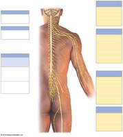
The spinal cord extends from the foramen magnum at the base of the skull to the level of the first or second lumbar vertebra (L1–L2). It is divided into four regions:
Cervical
Thoracic
Lumbar
Sacral
Key anatomical features include:
Cervical enlargement: Supplies nerves to the upper limbs.
Lumbosacral enlargement: Supplies nerves to the lower limbs.
Conus medullaris: The tapered end of the spinal cord, usually at L1–L2.
Filum terminale: Anchors the spinal cord to the coccyx.
Cauda equina: Bundle of nerve roots continuing below the cord's end.
Spinal Nerves and Nerve Plexuses
Spinal Nerve Organization
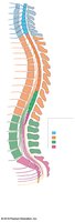
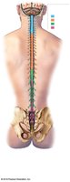
There are 31 pairs of spinal nerves, named for the segment of the spinal cord from which they emerge:
Cervical nerves: 8 pairs
Thoracic nerves: 12 pairs
Lumbar nerves: 5 pairs
Sacral nerves: 5 pairs
Coccygeal nerve: 1 pair
Spinal nerves are part of the peripheral nervous system (PNS) and are named and numbered according to their vertebral level.
Nerve Plexuses
Nerve plexuses are networks formed by the anterior rami of spinal nerves, containing both sensory and motor fibers. The four major plexuses are:
Cervical plexus (C1–C5): Innervates neck muscles and the diaphragm.
Brachial plexus (C5–T1): Innervates pectoral girdle and upper limbs.
Lumbar plexus (T12–L4): Innervates pelvic girdle and lower limbs.
Sacral plexus (L4–S4): Innervates pelvic girdle and lower limbs.
Internal Anatomy of the Spinal Cord
Gray and White Matter
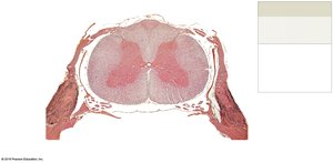
The spinal cord's internal structure consists of gray matter and white matter:
Gray matter: Contains cell bodies of neurons and glial cells; located centrally in the cord.
White matter: Contains myelinated axons; surrounds the gray matter.
Two grooves run the length of the cord:
Anterior median fissure: Deep and wide.
Posterior median sulcus: Shallow and narrow.
The central canal is a small space in the center, filled with cerebrospinal fluid.
Functional Organization
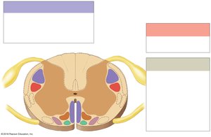
Posterior horn: Contains sensory nuclei.
Lateral horn: Contains visceral motor nuclei (in thoracic and upper lumbar regions).
Anterior horn: Contains somatic motor nuclei.
Gray commissure: Area where gray matter crosses the midline.
White matter is organized into columns and tracts:
Sensory tracts: Carry information toward the brain.
Motor tracts: Carry commands from the brain to effectors.
Spinal Nerve Structure and Coverings
Connective Tissue Layers
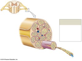
Spinal nerves are protected by three connective tissue layers:
Endoneurium: Surrounds individual axons.
Perineurium: Surrounds groups of axons (fascicles).
Epineurium: Surrounds the entire nerve.
Branches: Roots, Nerves, and Rami
Roots
Roots are the initial branches emerging from the spinal cord:
Dorsal (posterior) roots: Contain sensory fibers; associated with the dorsal root ganglion (cell bodies of sensory neurons).
Ventral (anterior) roots: Contain motor fibers.
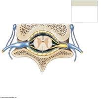
Spinal Nerves and Rami
Spinal nerves: Formed where dorsal and ventral roots meet; mixed nerves containing both sensory and motor fibers.
Rami: Branches off spinal nerves; contain both sensory and motor fibers. Posterior rami innervate muscles and skin of the back; anterior rami innervate limbs and anterior trunk.
Nerve Plexuses: Major Nerves and Distribution
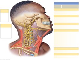
Cervical Plexus
The cervical plexus innervates muscles and skin of the neck and the diaphragm. The phrenic nerve controls the diaphragm, a major respiratory muscle. Severing the spinal cord above the origin of the phrenic nerve results in loss of diaphragmatic function.
Brachial Plexus
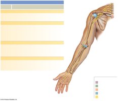
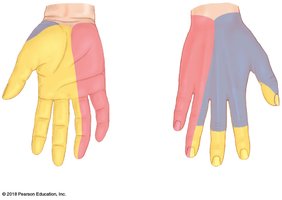
The brachial plexus gives rise to all nerves innervating the upper limb, including:
Musculocutaneous nerve
Radial nerve
Median nerve
Ulnar nerve
Lumbar and Sacral Plexuses
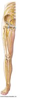
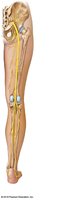
The lumbar plexus innervates the pelvic girdle and anterior/medial thigh. The sacral plexus includes nerves such as the sciatic, gluteal, and pudendal nerves, innervating the lower limb and pelvic region.
Dermatomes and Intercostal Nerves
Dermatomes
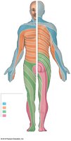
A dermatome is an area of skin innervated by a single spinal nerve. Dermatomes are clinically important for diagnosing nerve or spinal cord injuries.
Intercostal Nerves
Intercostal nerves (T1–T12) innervate the muscles and skin of the thoracic wall.
Spinal Cord Injuries
Types of Paralysis
Paraplegia: Spinal cord transected in the thoracic region; paralysis of lower limbs.
Quadriplegia: Spinal cord transected in the cervical region; paralysis of all four limbs.
Spinal Reflexes
Reflex Arc
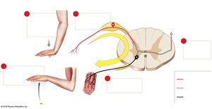
A reflex is a fast, involuntary response to a specific stimulus. The reflex arc is the pathway a nerve impulse takes to create a reflex, consisting of five components:
Stimulus activates receptor
Sensory neuron carries signal to CNS
Processing occurs in the CNS
Motor neuron carries signal to effector
Response by effector
Monosynaptic Reflexes
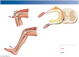
In monosynaptic reflexes, the sensory neuron directly synapses with the motor neuron. Example: Patellar (stretch) reflex, which prevents knee buckling while standing.
Polysynaptic Reflexes
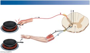
Polysynaptic reflexes involve one or more interneurons. Example: Withdrawal reflex, which causes automatic withdrawal from a painful stimulus.
Summary Table: Spinal Cord Regions and Nerves
Region | Number of Nerves | Main Functions |
|---|---|---|
Cervical | 8 pairs | Neck, upper limbs |
Thoracic | 12 pairs | Thorax, intercostal muscles |
Lumbar | 5 pairs | Lower limbs, pelvic girdle |
Sacral | 5 pairs | Lower limbs, pelvic organs |
Coccygeal | 1 pair | Pelvic region |
Key Terms and Definitions
Spinal cord: Central nervous system structure transmitting information between brain and body.
Meninges: Protective coverings of the CNS.
Spinal nerve: Mixed nerve emerging from the spinal cord.
Plexus: Network of nerves formed by anterior rami.
Dermatome: Skin area innervated by a single spinal nerve.
Reflex arc: Pathway mediating a reflex action.
Important Equations
While the spinal cord is primarily anatomical, the following formula is relevant for nerve conduction:
Nerve conduction velocity:
Where distance is the length the impulse travels, and time is the duration taken.
