 Back
BackStudy Notes: Introduction to the Musculoskeletal System
Study Guide - Smart Notes
 Tailored notes based on your materials, expanded with key definitions, examples, and context.
Musculoskeletal System
Overview
The musculoskeletal system is essential for movement, support, and protection of the body. It consists of bones, muscles, joints, and connective tissues, working together to maintain posture, produce movement, and protect internal organs.
Skeletal System
Functions of the Skeletal System
Support: Provides structural framework for the body.
Protection: Shields vital organs such as the brain, heart, and lungs.
Movement: Acts as levers for muscles to produce movement.
Storage: Stores minerals, especially calcium and phosphorus.
Blood Cell Formation: Houses bone marrow for hematopoiesis.
Composition and Function of Bone Tissue
Bones, or osseous tissue, are among the hardest materials in the body. They are formed through ossification, a process beginning before birth. The initial skeleton is made of cartilage, which is gradually replaced by bone. Bone tissue consists of specialized cells:
Osteoblasts: Build new bone tissue.
Osteoclasts: Break down bone tissue.
Osteocytes: Mature bone cells maintaining bone matrix.
Minerals such as calcium (Ca) and phosphorus (P) are crucial for strong bone formation.
Classification of Bones
The adult skeleton contains 206 bones, classified by shape:
Long Bones: e.g., femur, humerus
Flat Bones: e.g., skull, sternum
Irregular Bones: e.g., vertebrae
Short Bones: e.g., carpals, tarsals
Bone tissue types:
Compact Bone: Dense, hard outer layer.
Spongy Bone: Porous, inner layer.
Structure of the Bone
Diaphysis: Central shaft of long bones.
Epiphysis: Ends of long bones.
Periosteum: Thin connective tissue covering diaphysis.
Articular Cartilage: Covers epiphysis, reduces friction.
Bone Formation, Growth, and Remodeling
Bones grow and remodel throughout life, adapting to stress and repairing damage. Osteoblasts and osteoclasts play key roles in this dynamic process.
Fractures
Fractures are breaks in bone integrity, commonly resulting from trauma or pathological conditions. Types include simple, compound, comminuted, and greenstick fractures.
Axial Skeleton
Components
The axial skeleton forms the central axis of the body and includes:
Skull: Cranium (frontal, parietal, temporal, ethmoid, sphenoid, occipital) and facial bones (mandible, maxilla, zygomatic, vomer, palatine, nasal, lacrimal).
Vertebral Column: Extends from skull to pelvis, separated by intervertebral discs.
Ribs: 12 pairs, classified as true, false, and floating ribs.
Conditions of the Spine
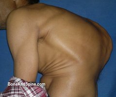
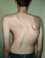
Abnormalities of the vertebral column can affect posture and function. Examples include scoliosis, kyphosis, and lordosis. These conditions may result in visible deformities and impact movement or organ function.
Ribs
True Ribs: First seven pairs, attach directly to sternum.
False Ribs: Next five pairs, attach indirectly or not at all.
Floating Ribs: Last two pairs of false ribs, no sternal attachment.
Intercostal Muscles: Fill spaces between ribs, aid in breathing.
Appendicular Skeleton
Components
Pectoral Girdle: Clavicle and scapula.
Upper Extremity: Humerus, ulna, radius, carpals, metacarpals, phalanges.
Pelvic Girdle: Os coxae (hipbone).
Lower Extremity: Femur, patella, tibia, fibula, tarsals, metatarsals, phalanges.
Joints
Overview
Joints are connections between bones, allowing movement and providing stability. Types include fibrous, cartilaginous, and synovial joints.
Muscular System
Functions of the Muscular System
Produces Movement: Muscles contract to move body parts.
Maintains Posture: Stabilizes body position.
Stabilizes Joints: Supports and protects joints.
Protects Internal Organs: Shields organs from injury.
Generates Heat: Muscle activity produces heat for temperature regulation.
Types of Muscles
Skeletal Muscle: Voluntary, striated, attached to bones, responsible for movement.
Smooth Muscle: Involuntary, non-striated, found in internal organs and blood vessels.
Cardiac Muscle: Involuntary, striated, found only in the heart.
Skeletal Muscle
Skeletal muscle fibers are wrapped in fascia, which tapers to form tendons. Movement is controlled by motor neurons at the myoneural junction. Skeletal muscle is voluntary and striated.
Smooth Muscle
Found in walls of hollow organs (e.g., stomach, blood vessels), smooth muscle is involuntary and responsible for actions such as peristalsis and vasoconstriction.
Cardiac Muscle
Cardiac muscle forms the heart wall (myocardium). It contracts involuntarily to pump blood throughout the body.
How Muscles Contract
Muscle contraction involves the interaction of actin and myosin filaments, powered by ATP. The process is initiated by nerve impulses and regulated by calcium ions.
Example: Muscle Contraction Equation
The sliding filament theory can be summarized as:
References
Tortora and Derrickson's Principles of Anatomy and Physiology (2021)
Ross and Wilson Anatomy and Physiology in Health and Illness (2022)
Human Anatomy & Physiology (Marieb & Hoehn, 2019)
National Health Service (2023) Musculoskeletal system information
