Back
BackStudy Notes: Joints (Articulations) in the Skeletal System
Study Guide - Smart Notes
 Tailored notes based on your materials, expanded with key definitions, examples, and context.
Joints (Articulations) in the Skeletal System
Introduction to Joints
Joints, or articulations, are sites where two or more bones meet. They play a crucial role in providing mobility to the skeleton while maintaining its structural integrity. Joints can also form between bone and cartilage or bone and teeth (gums).
Definition: A joint is the point of contact between bones, or between bone and cartilage/teeth.
Function: Joints allow for movement and flexibility, and some provide stability and support.
Classification of Joints by Movement
Joints are classified functionally based on the degree of movement they permit:
Synarthroses: Immovable joints (e.g., sutures of the skull).
Amphiarthroses: Slightly movable joints (e.g., intervertebral discs).
Diarthroses: Freely movable joints (e.g., most limb joints).
Structural Classification of Joints
Joints are also classified structurally based on the material binding the bones and the presence or absence of a joint cavity:
Fibrous Joints: Bones joined by dense fibrous connective tissue; no joint cavity; little or no movement.
Cartilaginous Joints: Bones joined by cartilage; no joint cavity; little or no movement.
Synovial Joints: Bones separated by a fluid-filled joint cavity; permit free movement.

Fibrous Joints
Types of Fibrous Joints
Fibrous joints are united by fibrous connective tissue and are generally immovable or allow very limited movement.
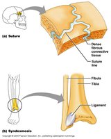
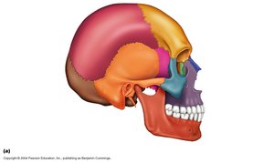
Sutures: Found only in the skull; bones interlock with a thin layer of dense connective tissue.
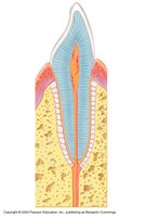
Gomphoses: Peg-in-socket joints, such as teeth in their sockets, held by periodontal ligaments.
Syndesmoses: Bones connected by a ligament or interosseous membrane; allow more movement than sutures but still limited (e.g., distal tibiofibular joint).

Features of Sutures
Located in the skull.
Articulating bones have a saw-tooth appearance.
Immovable (synarthroses).
Features of Gomphoses
Found between teeth and their sockets in the jaw.
Peg (tooth root) fits into a socket (alveolus) and is held by periodontal ligaments.
Immovable.
Features of Syndesmoses
Found between long bones such as the tibia and fibula.
Connected by a ligament or interosseous membrane.
Allows slight movement (amphiarthrotic).

Cartilaginous Joints
Types of Cartilaginous Joints
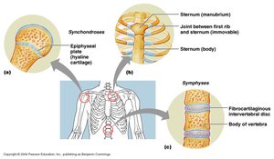
Symphyses: Bones joined by fibrocartilage; amphiarthrotic (slightly movable).
Synchondroses: Bones joined by hyaline cartilage; usually synarthrotic (immovable).

Features of Symphyses
Examples: Intervertebral discs, pubic symphysis.
Joint consists of a broad, flat disc of fibrocartilage stabilized by ligaments.
Allows slight movement (amphiarthrotic).

Intervertebral Disc Structure
Outer layer: Fibrocartilage (annulus fibrosus).
Inner core: Semi-liquid cartilage (nucleus pulposus).
Functions as a shock absorber and allows limited movement between vertebrae.

Features of Synchondroses
Examples: Epiphyseal plates in growing bones, joint between first rib and sternum.
Composed of hyaline cartilage.
Immovable (synarthroses).

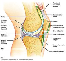
Synovial Joints
General Structure and Stabilizing Features
Synovial joints are the most common and movable type of joint in the body. They are characterized by the presence of a joint cavity filled with synovial fluid.
Bones are joined by a fibrous capsule (joint capsule) and ligaments.
Muscle arrangement and the interlocking fit of bones contribute to stability.
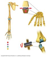
Types include hinge, pivot, and ball-and-socket joints.
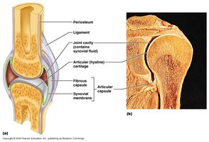
Fibrous Capsule
Surrounds the joint and is composed of dense connective tissue.
Provides strength and flexibility.
Joins with the periosteum of the bones.

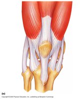
Ligaments
Thickenings of the joint capsule or separate bands of connective tissue.
Provide strength and limit excessive movement.
Sprains occur when ligament fibers are torn.
Additional Features of Synovial Joints
Synovial membrane lining the capsule produces synovial fluid for lubrication and nutrition.
Articular cartilage covers the ends of bones, reducing friction.
Menisci (pads of cartilage) act as shock absorbers and improve fit between bones.
Bursae are sacs filled with synovial fluid that reduce friction between tissues.
Fat pads cushion the joint.
Synovial Membrane
Composed of loose connective tissue.
Lines non-articular surfaces inside the joint capsule.
Produces synovial fluid for lubrication, nutrient distribution, and shock absorption.

Articular Cartilage
Hyaline cartilage covering the ends of bones in synovial joints.
Reduces friction and absorbs shock.
Receives nutrients from synovial fluid.

Menisci
Wedge-shaped pads of fibrocartilage within some synovial joints (e.g., knee).
Improve the fit between articulating bones, increase stability, and act as shock absorbers.

Bursae
Small sacs lined with synovial membrane and filled with synovial fluid.
Located between bones and muscles or bones and skin to reduce friction.

Fat Pads
Masses of adipose tissue within some synovial joints.
Act as cushions to protect the joint.

Joint Disorders
Dislocations
Occur when bones are forced out of their normal positions in a joint.
Common in the shoulder and fingers.
Osteoarthritis
Non-inflammatory degenerative joint disease.
Articular cartilage deteriorates, leading to the formation of bone spurs.
Typically affects large, weight-bearing joints first (e.g., knees, hips).
Associated with aging and wear-and-tear.
Rheumatoid Arthritis
Autoimmune disorder causing inflammation of the synovial membrane.
Leads to destruction of articular cartilage and joint deformity.
Usually affects small joints first (e.g., fingers, wrists).
May be managed by medications and dietary collagen supplementation.