 Back
BackStudy Notes: The Cardiovascular System – Heart and Great Vessels
Study Guide - Smart Notes
 Tailored notes based on your materials, expanded with key definitions, examples, and context.
Cardiovascular System
Overview
The cardiovascular system is responsible for the circulation of blood throughout the body, delivering oxygen and nutrients to tissues and removing waste products. The heart, a muscular organ, acts as the central pump, while the great vessels facilitate blood flow to and from the heart.
Heart and Great Vessels
External Anatomy of the Heart
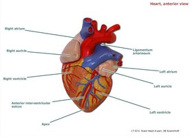
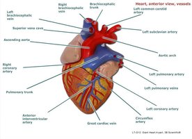
The heart is divided into four chambers: right atrium, left atrium, right ventricle, and left ventricle. Each chamber has distinct anatomical features and functions.
Right Atrium: Receives deoxygenated blood from the body via the superior and inferior vena cava and the coronary sinus.
Right Auricle: A small muscular pouch attached to the right atrium, increasing its capacity.
Left Atrium: Receives oxygenated blood from the lungs via the pulmonary veins.
Left Auricle: A muscular pouch attached to the left atrium.
Right Ventricle: Pumps deoxygenated blood to the lungs through the pulmonary trunk and arteries.
Left Ventricle: Pumps oxygenated blood to the systemic circulation via the aorta.
Ligamentum Arteriosum: A remnant of the fetal ductus arteriosus, connecting the pulmonary artery to the aorta.
Internal Anatomy of the Heart
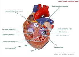
Internally, the heart contains valves and muscular structures that ensure unidirectional blood flow and prevent backflow.
Atrioventricular (AV) Valves:
Tricuspid Valve: Between right atrium and right ventricle.
Bicuspid/Mitral Valve: Between left atrium and left ventricle.
Semilunar Valves:
Pulmonary Semilunar Valve: Between right ventricle and pulmonary trunk.
Aortic Semilunar Valve: Between left ventricle and aorta.
Chordae Tendineae: Fibrous cords that anchor the AV valves to papillary muscles.
Papillary Muscles: Contract to prevent valve prolapse during ventricular contraction.
Interventricular Septum: Muscular wall separating the right and left ventricles.
Major Blood Vessels Associated with the Heart
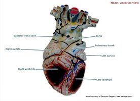
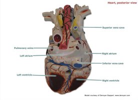
The heart is connected to several major vessels that transport blood to and from the organ.
Superior and Inferior Vena Cava: Bring deoxygenated blood from the body to the right atrium.
Pulmonary Veins: Carry oxygenated blood from the lungs to the left atrium.
Pulmonary Trunk and Arteries: Carry deoxygenated blood from the right ventricle to the lungs.
Aorta: Distributes oxygenated blood from the left ventricle to the systemic circulation.
Coronary Arteries: Supply blood to the myocardium (heart muscle).
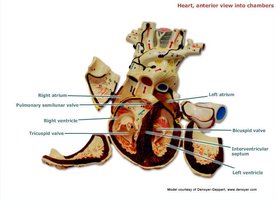
Heart Chambers and Valves (Sectional View)
Sectional views of the heart reveal the arrangement of chambers and valves, crucial for understanding blood flow.
Right Atrium and Ventricle: Tricuspid valve, pulmonary semilunar valve.
Left Atrium and Ventricle: Bicuspid/mitral valve, aortic semilunar valve.
Interventricular Septum: Separates the ventricles.
Systemic Circulation
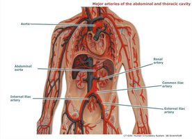
Branches of the Aorta
The aorta is the main artery of the body, branching to supply blood to various regions.
To the Myocardium:
Left coronary artery
Right coronary artery
To the Head/Neck and Upper Limbs:
Brachiocephalic trunk
Common carotid arteries
Subclavian arteries
To the Abdomen and Lower Limbs:
Celiac trunk
Superior mesenteric artery
Inferior mesenteric artery
Common iliac arteries
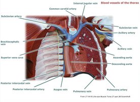
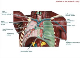
Blood Vessels of the Thorax
The thoracic cavity contains several important arteries and veins that supply the heart and lungs.
Arteries: Subclavian, common carotid, aortic arch, pulmonary artery, posterior intercostal arteries.
Veins: Subclavian, brachiocephalic, superior vena cava, pulmonary veins, azygos vein.
Cardiovascular System Histology
Histology of Blood Vessels
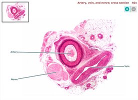
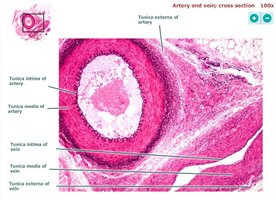
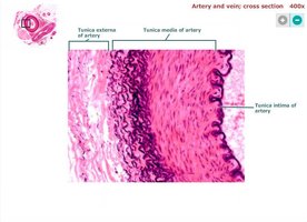
Blood vessels are composed of three main layers: tunica intima, tunica media, and tunica externa. These layers differ between arteries and veins.
Tunica Intima: Innermost layer, composed of endothelial cells.
Tunica Media: Middle layer, primarily smooth muscle (thicker in arteries).
Tunica Externa: Outermost layer, connective tissue.
Arteries vs. Veins: Arteries have thicker tunica media, veins have larger lumens and thinner walls.
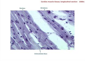
Histology of Cardiac Muscle Tissue
Cardiac muscle tissue is unique to the heart, characterized by striations, intercalated discs, and centrally located nuclei.
Striations: Alternating light and dark bands due to sarcomere structure.
Intercalated Discs: Specialized junctions that allow rapid electrical communication between cells.
Nucleus: Usually one per cell, centrally located.
Summary Table: Heart Chambers and Associated Structures
Chamber | Main Vessels | Valves | Key Features |
|---|---|---|---|
Right Atrium | Superior/Inferior Vena Cava, Coronary Sinus | Tricuspid Valve | Right Auricle, Fossa Ovalis |
Left Atrium | Pulmonary Veins | Bicuspid/Mitral Valve | Left Auricle |
Right Ventricle | Pulmonary Trunk | Pulmonary Semilunar Valve | Chordae Tendineae, Papillary Muscles |
Left Ventricle | Aorta | Aortic Semilunar Valve | Chordae Tendineae, Papillary Muscles |
Key Terms and Definitions
Myocardium: The muscular layer of the heart responsible for contraction.
Coronary Arteries: Vessels that supply blood to the heart muscle.
Interventricular Septum: Wall separating the right and left ventricles.
Chordae Tendineae: Tendinous cords anchoring AV valves to papillary muscles.
Ligamentum Arteriosum: Remnant of fetal circulation connecting pulmonary artery and aorta.
Example: Blood Flow Through the Heart
Blood enters the right atrium from the body, passes through the tricuspid valve to the right ventricle, and is pumped to the lungs via the pulmonary semilunar valve. Oxygenated blood returns to the left atrium, passes through the bicuspid valve to the left ventricle, and is pumped to the body via the aortic semilunar valve.
Relevant Equations
Blood pressure is determined by cardiac output and peripheral resistance:
Where:
BP: Blood Pressure
CO: Cardiac Output
PR: Peripheral Resistance
Cardiac output is calculated as:
Where:
HR: Heart Rate
SV: Stroke Volume
Additional info: Academic context and expanded explanations have been added to ensure completeness and clarity for exam preparation.
