 Back
BackStudy Notes: The Nervous System (Anatomy & Physiology)
Study Guide - Smart Notes
 Tailored notes based on your materials, expanded with key definitions, examples, and context.
The Nervous System
Overview and Functions
The nervous system is a complex network responsible for coordinating the body's activities by transmitting signals between different parts. It is essential for sensation, movement, cognition, and homeostasis.
Key Functions: Sensory input, integration of information, motor output, regulation of homeostasis.
Major Divisions: Central Nervous System (CNS) and Peripheral Nervous System (PNS).
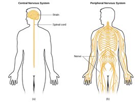
Organization of the Nervous System
Central vs. Peripheral Nervous System
The nervous system is divided into the CNS and PNS, each with distinct structures and functions.
CNS: Composed of the brain and spinal cord; main control center.
PNS: Composed of nerves outside the CNS; connects CNS to limbs and organs.
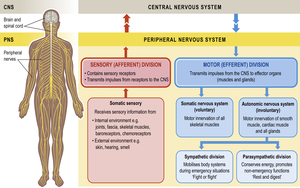
Functional Divisions of the PNS
Sensory (Afferent) Division: Transmits sensory information from receptors to the CNS.
Motor (Efferent) Division: Transmits impulses from CNS to effectors (muscles and glands).
Somatic Nervous System: Controls voluntary movements of skeletal muscles.
Autonomic Nervous System: Controls involuntary functions (e.g., heart rate, digestion).
Sympathetic Division: Prepares body for 'fight or flight'.
Parasympathetic Division: Promotes 'rest and digest'.
Cells and Tissues of the Nervous System
Neuroglia (Glial Cells)
Neuroglia are supporting cells that protect, nourish, and maintain neurons. They do not transmit impulses but are essential for neural health and function.
Types: Astrocytes, oligodendrocytes, microglia, Schwann cells, ependymal cells.
Functions: Support, insulation, immune defense, and maintenance of the extracellular environment.
Neurones (Neurons)
Neurons are specialized cells that carry electrical impulses. They are the fundamental units of the nervous system.
Structure: Cell body, dendrites, axon, axon terminals.
Function: Generate and transmit action potentials in response to stimuli.
Bundles of axons: Called nerves in the PNS.
Cell bodies: Form grey matter; grouped as nuclei (CNS) or ganglia (PNS).
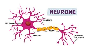
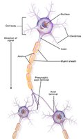
Axons and Dendrites
Axons: Single, long extension; carries impulses away from cell body.
Dendrites: Multiple, branched extensions; receive signals and carry them toward cell body.
White matter: Bundles of myelinated axons.
Myelination
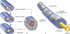
Myelin sheath is a fatty layer that insulates axons, increasing the speed of impulse transmission.
Schwann cells: Form myelin in PNS.
Oligodendrocytes: Form myelin in CNS.
Nodes of Ranvier: Gaps in myelin sheath where action potentials are regenerated.
Action Potential (Nerve Impulse)
Generation and Transmission
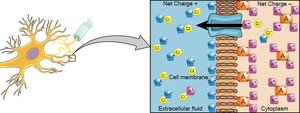
An action potential is an electrical signal generated by the movement of ions across the neuron's membrane. It is the fundamental mechanism for communication in the nervous system.
Initiation: Triggered by stimulation of sensory nerve endings or another nerve.
Transmission: Involves movement of sodium (Na+) and potassium (K+) ions.
Resting potential: Maintained by sodium-potassium pump.
Depolarization: Na+ influx causes membrane potential to become positive.
Repolarization: K+ efflux restores negative membrane potential.
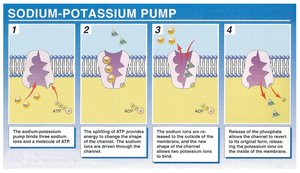
Equation: The sodium-potassium pump exchanges three sodium ions for two potassium ions, using ATP:
The Synapse and Neurotransmitters
Synaptic Transmission
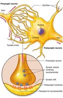
A synapse is the junction between two neurons where signals are transmitted chemically via neurotransmitters. There is no physical contact between neurons; the signal crosses a small gap called the synaptic cleft.
Presynaptic neuron: Releases neurotransmitter.
Postsynaptic neuron: Receives neurotransmitter.
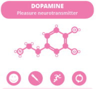
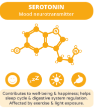
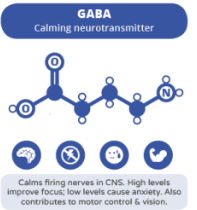
Neurotransmitters: Chemical messengers (e.g., dopamine, serotonin, GABA).
Major Neurotransmitters and Their Functions
Dopamine: Pleasure, reward, motor control.
Serotonin: Mood, sleep, appetite.
GABA: Calming, inhibitory effects.
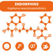
Endorphins: Euphoria, pain relief.
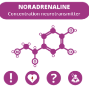
Noradrenaline: Concentration, alertness.
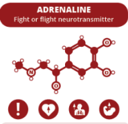
Adrenaline: Fight or flight response.
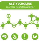
Acetylcholine: Learning, memory, muscle contraction.
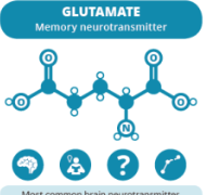
Glutamate: Excitatory, memory, learning.
Nerves and Sensory Receptors
Types of Nerves
Sensory (Afferent) Nerves: Carry information from receptors to CNS.
Motor (Efferent) Nerves: Carry commands from CNS to muscles and glands.
Autonomic Nerves: Control involuntary functions.
Central Nervous System (CNS)
Structure and Protection
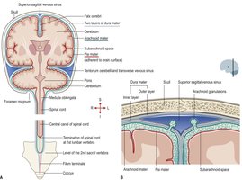
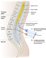
The CNS consists of the brain and spinal cord, protected by the skull, vertebrae, and three layers of meninges.
Dura mater: Tough, outermost layer.
Arachnoid mater: Middle, web-like layer.
Pia mater: Delicate, innermost layer.
Spaces: Subdural (between dura and arachnoid), subarachnoid (between arachnoid and pia, contains CSF), epidural (spinal canal).
Brain Structure and Lobes
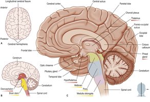
The brain is divided into several major parts and lobes, each with specialized functions.
Cerebrum: Higher functions, sensory processing, voluntary movement.
Thalamus: Relay center for sensory information.
Hypothalamus: Homeostasis, hormone regulation.
Midbrain, Pons, Medulla oblongata: Brainstem functions, vital processes.
Brain Lobes and Their Functions
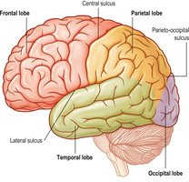
Frontal lobe: Movement, higher thinking, personality, speech production (Broca's area).
Parietal lobe: Sensory interpretation, spatial relationships, attention, language.
Temporal lobe: Processing smell, taste, sound, memory, language, reading.
Occipital lobe: Vision, visual processing, recognition of shapes and colors.
Brain Hemispheres
Left and Right Hemispheres
The brain is composed of two hemispheres connected by the corpus callosum. Each hemisphere controls the opposite side of the body and has specialized functions.
Corpus callosum: Facilitates communication between hemispheres.
Contralateral control: Each hemisphere controls the opposite side of the body.
Specialization: Right hemisphere: spatial, artistic, facial perception; Left hemisphere: language, logic.
Ventricles and Cerebrospinal Fluid (CSF)
Brain Ventricles
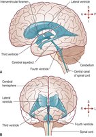
The brain contains four ventricles filled with CSF: right and left lateral ventricles, third ventricle, and fourth ventricle.
Functions of CSF
Shock absorption: Protects brain from injury.
Nutrition: Supplies essential nutrients.
Intracranial pressure: Maintains stable pressure.
Waste removal: Cleanses toxins and waste.
Temperature regulation: Keeps brain and spine temperature stable.
Immune function: Contains immune cells for defense.
Reflex Arc
Mechanism of Reflexes
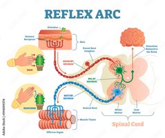
A reflex arc is the pathway by which a reflex action occurs, involving sensory input, integration in the spinal cord, and motor output.
Sensory receptor: Detects stimulus.
Sensory neuron: Transmits signal to CNS.
Relay neuron: Integrates information in spinal cord.
Motor neuron: Sends command to effector (muscle).
Effector: Produces response (e.g., withdrawal from pain).
Summary Table: Major Neurotransmitters
Neurotransmitter | Main Function | Example/Application |
|---|---|---|
Dopamine | Pleasure, reward, motor control | Parkinson's disease, addiction |
Serotonin | Mood, sleep, appetite | Depression, anxiety |
GABA | Calming, inhibitory | Epilepsy, anxiety |
Endorphins | Euphoria, pain relief | Runner's high, pain management |
Noradrenaline | Concentration, alertness | Attention, stress response |
Adrenaline | Fight or flight | Acute stress, emergency response |
Acetylcholine | Learning, memory, muscle contraction | Alzheimer's disease, muscle function |
Glutamate | Excitatory, memory, learning | Neuroplasticity, stroke |
Additional info: These notes are based on standard anatomy and physiology textbooks and lecture materials, with expanded academic context for completeness.
