 Back
BackStudy Notes: The Nervous System – Structure, Function, and Organization
Study Guide - Smart Notes
 Tailored notes based on your materials, expanded with key definitions, examples, and context.
The Nervous System: Structure, Function, and Organization
Overview of the Nervous System
The nervous system is a complex network responsible for coordinating the body's activities by transmitting signals to and from different parts of the body. It is divided into the central nervous system (CNS) and the peripheral nervous system (PNS). The CNS includes the brain and spinal cord, while the PNS consists of nerves and ganglia outside the CNS.
Peripheral Nervous System (PNS)
Organization of Peripheral Nerves
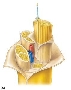
Peripheral nerves are bundles of axons that transmit sensory and motor information between the CNS and the rest of the body. Each nerve is composed of several layers of connective tissue:
Endoneurium: Surrounds individual axons.
Perineurium: Encloses bundles of axons (fascicles).
Epineurium: Outermost layer, encasing the entire nerve.
Blood vessels: Supply nutrients and oxygen to nerve fibers.
Brachial Plexus and Nerves of the Arm
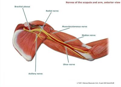
The brachial plexus is a network of nerves that originates from the spinal cord in the neck and supplies the shoulder, arm, and hand. Major nerves arising from the brachial plexus include:
Axillary nerve: Innervates the deltoid and teres minor muscles.
Radial nerve: Supplies the posterior compartment of the arm and forearm.
Musculocutaneous nerve: Innervates the anterior compartment of the arm.
Median nerve: Supplies most of the anterior forearm and some hand muscles.
Ulnar nerve: Innervates muscles of the hand and some forearm muscles.
Sacral Plexus and Lower Limb Innervation
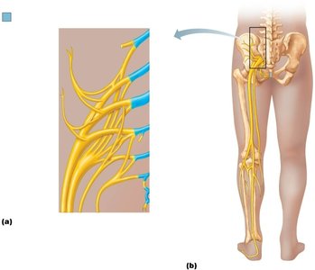
The sacral plexus is formed by the ventral rami of spinal nerves L4-S4 and supplies the pelvis, buttocks, genitals, thighs, calves, and feet. The largest nerve from this plexus is the sciatic nerve, which branches into the tibial and common fibular nerves.
Neurons and Synapses
Structure of a Myelinated Neuron
Neurons are the functional units of the nervous system. A typical myelinated neuron consists of:
Cell body (soma): Contains the nucleus and organelles.
Dendrites: Receive incoming signals.
Axon: Conducts electrical impulses away from the cell body.
Myelin sheath: Insulating layer formed by Schwann cells in the PNS, which increases the speed of impulse conduction.
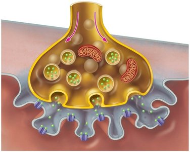
The Neuromuscular Junction
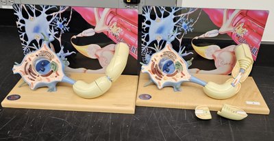
The neuromuscular junction is a specialized synapse where a motor neuron communicates with a skeletal muscle fiber. Key features include:
Axon terminal: Contains synaptic vesicles filled with acetylcholine (ACh).
Synaptic cleft: The gap between the neuron and muscle fiber.
Junctional folds: Increase surface area for ACh receptors on the muscle cell membrane (sarcolemma).
Signal transmission: Release of ACh triggers muscle contraction.
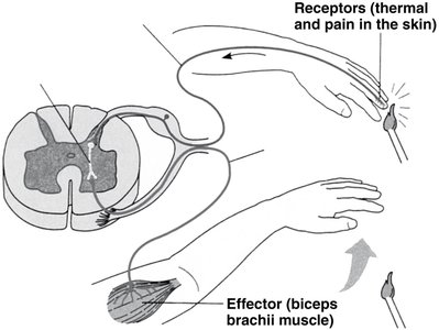
Reflex Arcs
Components of a Reflex Arc
A reflex arc is the neural pathway that mediates a reflex action. It typically involves:
Receptor: Detects a stimulus (e.g., pain, temperature).
Sensory neuron: Transmits the signal to the CNS.
Integration center: Usually within the spinal cord.
Motor neuron: Carries the response signal to the effector.
Effector: Muscle or gland that carries out the response.
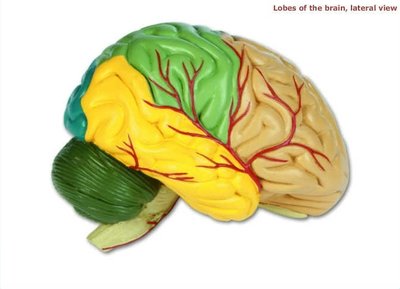
Brain Structure and Organization
Lobes and Major Regions of the Brain
The brain is divided into several lobes, each associated with specific functions:
Frontal lobe: Involved in reasoning, planning, movement, and problem-solving.
Parietal lobe: Processes sensory information such as touch, temperature, and pain.
Temporal lobe: Responsible for hearing and memory.
Occipital lobe: Main center for visual processing.
Cerebellum: Coordinates voluntary movements and balance.
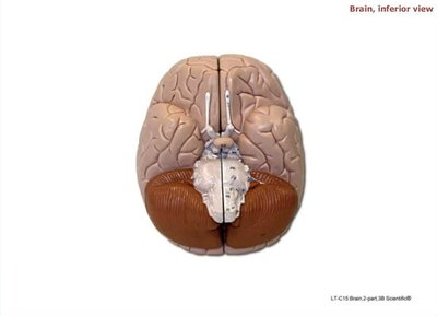
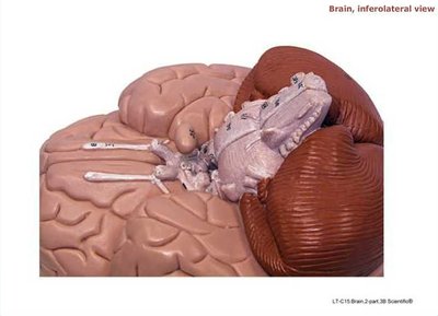
Inferior and Inferolateral Views of the Brain
Viewing the brain from below (inferior view) or from an inferolateral angle reveals the cranial nerves and the cerebellum. The cranial nerves are numbered I-XII and serve sensory, motor, or mixed functions.
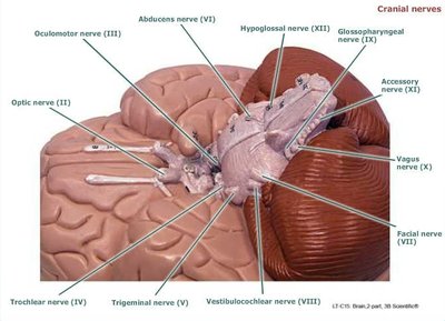
Cranial Nerves
The twelve pairs of cranial nerves emerge from the brain and brainstem, each with specific sensory and/or motor functions. Examples include:
Olfactory nerve (I): Sense of smell.
Optic nerve (II): Vision.
Oculomotor nerve (III): Eye movement.
Facial nerve (VII): Facial expression and taste.
Vagus nerve (X): Parasympathetic control of the heart, lungs, and digestive tract.
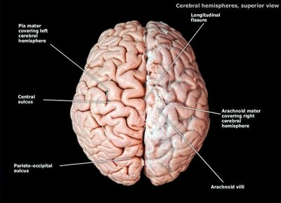
Major Sulci and Gyri
The surface of the cerebral hemispheres is marked by grooves (sulci) and ridges (gyri) that increase the brain's surface area and allow for greater cognitive function. Important sulci include the central sulcus and parieto-occipital sulcus.
Brain Ventricles
The brain contains interconnected cavities called ventricles, which are filled with cerebrospinal fluid (CSF). The main ventricles are:
Lateral ventricles (right and left)
Third ventricle
Fourth ventricle
Summary Table: Major Nerves of the Upper Limb
Nerve | Origin | Innervated Muscles/Regions |
|---|---|---|
Axillary | Brachial plexus (C5-C6) | Deltoid, teres minor |
Radial | Brachial plexus (C5-T1) | Posterior arm and forearm |
Musculocutaneous | Brachial plexus (C5-C7) | Anterior arm |
Median | Brachial plexus (C5-T1) | Anterior forearm, some hand muscles |
Ulnar | Brachial plexus (C8-T1) | Hand muscles, some forearm muscles |
Key Terms and Definitions
Plexus: A network of intersecting nerves.
Myelin sheath: Fatty covering that speeds up nerve impulse transmission.
Synapse: Junction between two neurons or a neuron and its target cell.
Reflex arc: Neural pathway that controls a reflex action.
Cranial nerves: Twelve pairs of nerves that emerge directly from the brain.
