 Back
BackStudy Notes: The Nervous System – Structure, Function, and Anatomy
Study Guide - Smart Notes
 Tailored notes based on your materials, expanded with key definitions, examples, and context.
The Nervous System
Structural and Functional Classification
The nervous system is classified both structurally and functionally to better understand its organization and roles in the human body.
Structural Classification:
Central Nervous System (CNS): Consists of the brain and spinal cord. Responsible for integrating, processing, and coordinating sensory data and motor commands.
Peripheral Nervous System (PNS): Includes all neural tissue outside the CNS. Divided into cranial nerves, spinal nerves, and plexuses.
Functional Classification:
Sensory (Afferent) Division: Transmits sensory information from receptors to the CNS.
Motor (Efferent) Division: Carries commands from the CNS to effectors (muscles and glands). Subdivided into:
Somatic Nervous System: Controls voluntary movements of skeletal muscles.
Autonomic Nervous System: Regulates involuntary functions (e.g., heart rate, digestion). Includes sympathetic and parasympathetic divisions.
Neuron Anatomy and Types of Cells in the Nervous System
Neuron Structure and Function
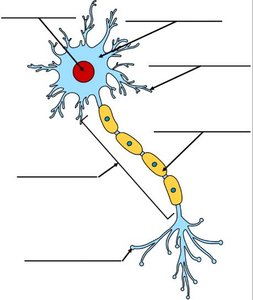
Neurons are the fundamental units of the nervous system, specialized for transmitting electrical impulses. They consist of several distinct parts, each with a specific function.
Dendrites: Receive incoming signals from other neurons.
Cell Body (Soma): Contains the nucleus and organelles; integrates incoming signals.
Axon: Conducts electrical impulses away from the cell body.
Myelin Sheath: Insulates the axon, speeding up impulse transmission.
Nodes of Ranvier: Gaps in the myelin sheath where action potentials are regenerated.
Axon Terminals: Release neurotransmitters to communicate with other neurons or effectors.
Types of Cells in the Nervous System
Neurons: Transmit electrical signals.
Glial Cells: Support and protect neurons. Types include:
Astrocytes: Maintain the blood-brain barrier and provide structural support.
Oligodendrocytes: Form myelin in the CNS.
Schwann Cells: Form myelin in the PNS.
Microglia: Act as immune cells in the CNS.
Ependymal Cells: Line ventricles and produce cerebrospinal fluid.
Nerve Transmission – Action Potential
Mechanism of Action Potential
Nerve transmission relies on the generation and propagation of action potentials, which are rapid changes in membrane potential that travel along the axon.
Resting Potential: The neuron maintains a negative charge inside relative to outside, typically around -70 mV.
Depolarization: Sodium channels open, allowing Na+ ions to enter, making the inside more positive.
Repolarization: Potassium channels open, K+ ions exit, restoring the negative charge.
Hyperpolarization: The membrane potential temporarily becomes more negative than the resting potential.
Propagation: The action potential travels down the axon, jumping between nodes of Ranvier (saltatory conduction).
Equation:
Example: When you touch a hot object, sensory neurons transmit the signal to the CNS, which processes it and sends a motor command to withdraw your hand.
CNS Structures
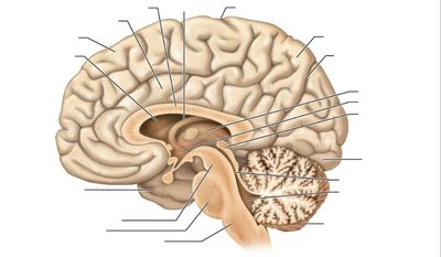
Major Structures of the Central Nervous System
The CNS is composed of the brain and spinal cord, each with specialized regions responsible for distinct functions.
Cerebrum: Controls higher functions such as reasoning, memory, and voluntary movement.
Cerebellum: Coordinates movement and balance.
Brainstem: Regulates vital functions like breathing and heart rate.
Thalamus: Relays sensory information to the cerebrum.
Hypothalamus: Maintains homeostasis and controls the endocrine system.
Spinal Cord: Conducts signals between the brain and the rest of the body; mediates reflexes.
PNS: Cranial Nerves, Spinal Nerves, and Plexuses
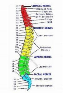
Peripheral Nervous System Organization
The PNS connects the CNS to limbs and organs, facilitating communication throughout the body.
Cranial Nerves: 12 pairs, primarily serving the head and neck.
Spinal Nerves: 31 pairs, emerging from the spinal cord and serving the rest of the body.
Plexuses: Networks of nerves formed by spinal nerves; include cervical, brachial, lumbar, and sacral plexuses.
Sympathetic and Parasympathetic Systems
Autonomic Nervous System Divisions
The autonomic nervous system regulates involuntary functions and is divided into two main branches:
Sympathetic System: Prepares the body for 'fight or flight' responses. Increases heart rate, dilates pupils, inhibits digestion.
Parasympathetic System: Promotes 'rest and digest' activities. Decreases heart rate, constricts pupils, stimulates digestion.
Comparison Table:
Function | Sympathetic | Parasympathetic |
|---|---|---|
Heart Rate | Increases | Decreases |
Pupil | Dilates | Constricts |
Digestion | Inhibits | Stimulates |
Respiration | Increases | Decreases |
Cranial Nerves: Sensory, Motor, and Mixed
Overview of Cranial Nerves
Cranial nerves are classified based on their function as sensory, motor, or mixed. Each nerve has a specific role in transmitting information to and from the brain.
Sensory Nerves: Carry sensory information (e.g., olfactory, optic).
Motor Nerves: Control muscle movements (e.g., oculomotor, abducens).
Mixed Nerves: Have both sensory and motor functions (e.g., trigeminal, facial).
Example: The facial nerve (VII) is a mixed nerve, controlling facial expressions and transmitting taste sensations.
Additional info: For detailed labeling and functions, refer to the provided diagrams and tables.
