 Back
BackCh 13 &14: The Spinal Cord and Brain (Chapters 13 & 14) – Structure and Function
Study Guide - Smart Notes
 Tailored notes based on your materials, expanded with key definitions, examples, and context.
The Spinal Cord
Gross Anatomy of the Spinal Cord
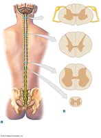
The spinal cord is a major component of the central nervous system, serving as a conduit for sensory and motor information between the body and brain. It is organized into distinct regions and contains both white and gray matter.
White Matter: Composed of myelinated and unmyelinated axons, located superficially, and organized into columns (funiculi) that carry ascending (sensory) and descending (motor) information.
Gray Matter: Contains neuron cell bodies, neuroglia, and unmyelinated axons. It surrounds the central canal and is organized into horns (anterior, lateral, posterior).
Key Landmarks: Anterior median fissure, posterior median sulcus, conus medullaris, filum terminale, central canal, dorsal and ventral roots, cauda equina.
White Matter vs. Gray Matter
White and gray matter are distributed differently along the spinal cord, with the proportion of white matter decreasing as you descend due to the exit of ascending fibers and entry of descending fibers.
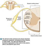
White Matter: Contains tracts of axons; organized into posterior, lateral, and anterior columns.
Gray Matter: Contains nuclei for sensory (dorsal) and motor (ventral) functions.
Dorsal and Ventral Roots
The spinal nerves are formed by the union of dorsal (sensory) and ventral (motor) roots. The dorsal root contains sensory axons and has an associated dorsal root ganglion, while the ventral root contains motor axons.
Dorsal Root: Sensory input to the spinal cord.
Ventral Root: Motor output from the spinal cord.
Meninges of the Spinal Cord
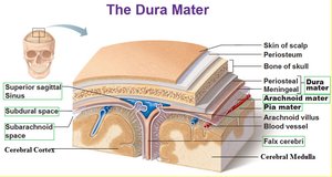
The spinal cord is protected by three connective tissue layers called meninges, which are continuous with those of the brain.
Dura Mater: Outermost, tough layer; separated from the vertebrae by the epidural space.
Arachnoid Mater: Middle layer; separated from the dura by the subdural space and from the pia by the subarachnoid space (contains cerebrospinal fluid).
Pia Mater: Innermost, delicate layer; adheres to the surface of the spinal cord.
Organization of Gray Matter
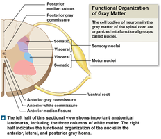
Gray matter is organized into nuclei, which are clusters of neuron cell bodies with specific functions.
Sensory Nuclei: Located dorsally; receive and process sensory information from peripheral receptors.
Motor Nuclei: Located ventrally; send motor commands to effectors (muscles and glands).
Somatic vs. Visceral: Somatic nuclei control skeletal muscles; visceral nuclei control smooth muscle, cardiac muscle, and glands.
Spinal Reflexes
Spinal reflexes are automatic, rapid responses to stimuli that do not require conscious thought. They are essential for protection and maintaining posture.
Monosynaptic Reflex: Involves one synapse (e.g., knee-jerk reflex).
Polysynaptic Reflex: Involves multiple synapses (e.g., withdrawal from a hot object).
Conditional Reflexes: Acquired through experience and memory.
The Brain
Major Regions of the Brain
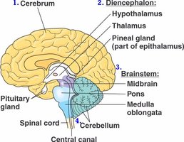
The brain is divided into four main regions, each with specialized functions:
Cerebrum: Largest part; responsible for higher mental functions, voluntary movement, and sensory processing.
Cerebellum: Coordinates movement and balance.
Diencephalon: Includes the thalamus and hypothalamus; relays sensory information and regulates autonomic functions.
Brainstem: Includes the midbrain, pons, and medulla oblongata; controls vital functions and connects the brain to the spinal cord.
The Cerebrum
The cerebrum is divided into left and right hemispheres, each with a surface layer of gray matter (cerebral cortex) that is highly folded to increase surface area.
Gyri: Elevated ridges.
Sulci: Shallow grooves.
Fissures: Deep grooves separating major regions.
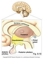
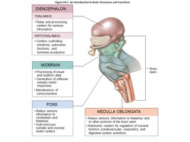
The Diencephalon
The diencephalon is located beneath the cerebrum and consists of the thalamus, hypothalamus, and epithalamus (including the pineal gland).
Thalamus: Relay and processing center for sensory information.
Hypothalamus: Regulates hormone production, emotions, autonomic functions, and homeostasis.
The Brainstem
The brainstem connects the cerebrum and cerebellum to the spinal cord and is responsible for many automatic functions necessary for survival.
Midbrain: Processes visual and auditory information; controls reflexes.
Pons: Relays sensory information and helps regulate breathing.
Medulla Oblongata: Controls autonomic functions such as heart rate, blood pressure, and digestion.
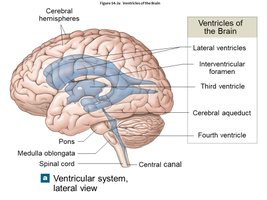
Ventricles of the Brain
The brain contains interconnected cavities called ventricles, which are filled with cerebrospinal fluid (CSF) and lined with ependymal cells.
Lateral Ventricles: One in each cerebral hemisphere; separated by the septum pellucidum.
Third Ventricle: Located in the diencephalon; communicates with lateral ventricles via the interventricular foramen.
Fourth Ventricle: Located between the brainstem and cerebellum; connects to the central canal of the spinal cord.
Protection and Support of the Brain
The brain is protected by the skull, cranial meninges, and cerebrospinal fluid. The blood-brain barrier provides biochemical isolation.
Cranial Meninges: Dura mater, arachnoid mater, and pia mater.
Cerebrospinal Fluid (CSF): Cushions the brain, provides nutrients, and removes waste; produced by the choroid plexus.
Blood-Brain Barrier: Formed by tight junctions between endothelial cells; restricts passage of substances from blood to brain tissue.
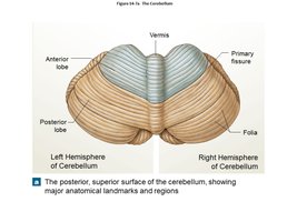
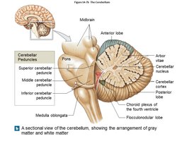
The Cerebellum
The cerebellum is responsible for coordinating voluntary movements, maintaining posture, and balance. It consists of two hemispheres and three lobes (anterior, posterior, flocculonodular).
Arbor Vitae: Tree-like arrangement of white matter.
Cerebellar Peduncles: Superior, middle, and inferior tracts connecting the cerebellum to the brainstem.
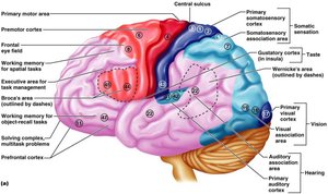
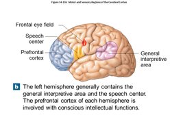
Functional Areas of the Cerebrum
Different regions of the cerebral cortex are specialized for various sensory, motor, and integrative functions.
Frontal Eye Field: Controls learned eye movements.
Broca’s Area: Speech production and regulation of breathing/vocalization.
Prefrontal Cortex: Intellectual functions, decision-making, and predicting consequences.
Wernicke’s Area: Language comprehension and analytical processing.
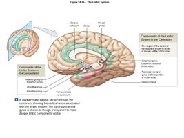
The Limbic System
The limbic system is a functional grouping of structures involved in emotion, memory, and motivation. It links conscious and unconscious brain functions.
Key Structures: Diencephalon, fornix, cingulate gyrus, parahippocampal gyrus, hippocampus.
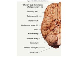
Cranial Nerves
There are 12 pairs of cranial nerves, each with specific sensory, motor, or mixed functions. They innervate structures in the head and neck, and some extend to thoracic and abdominal organs.
Sensory Nerves: Carry somatic sensory information (touch, pressure, vibration, temperature, pain).
Special Sensory Nerves: Carry sensations such as smell, sight, hearing, and balance.
Motor Nerves: Axons of somatic motor neurons.
Mixed Nerves: Contain both sensory and motor fibers.
Summary Table: Key Features of the Spinal Cord and Brain
Structure | Main Function | Key Features |
|---|---|---|
Spinal Cord | Conduction of sensory and motor information; reflexes | White and gray matter, dorsal/ventral roots, meninges |
Cerebrum | Higher mental functions, voluntary movement, sensory processing | Gyri, sulci, fissures, functional areas |
Cerebellum | Coordination of movement, balance | Arbor vitae, peduncles, lobes |
Diencephalon | Sensory relay, autonomic regulation, hormone production | Thalamus, hypothalamus, pineal gland |
Brainstem | Vital autonomic functions, pathway between brain and spinal cord | Midbrain, pons, medulla oblongata |
Cranial Nerves | Sensory and motor innervation of head/neck | 12 pairs, classified by function |
